Showing posts with label ACIS. Show all posts

Saturday, April 14, 2012

Wound Neovascularization with Dermal Substitutes

April 14, 2012

This is the second of 2 articles detailing wound healing studies done in nude mice implanted with different biological materials.  Full thickness skin wounds were created on the dorsum and then different dermal replacements were implanted.  Healing was followed for 28 days and very detailed histological and immunostaining evaluations of the wounds were performed to quantify the healing process.  Most studies of wound healing use simple or highly subjective measures of healing, but one of the main goals of these studies was to use objective criteria to quantify healing and to do so in fine detail.

Below is the full text of this article and also a PDF link of higher quality.


Wound Neovascularization and Dermal Substitutes in Nude Mice


Neovascularization of Wounds Treated with Dermal Substitutes in Nude Mice

by


Michele M. Loor, MD, Anh-Tuan Truong, MD, Barbara A. Latenser, MD 1,2,3 ,

Dorion E. Wiley, MD 1,2, and Robert J. Walter, PhD 1,2


1 Sumner L. Koch Burn Center, Department of Trauma, John Stroger Jr. Hospital of Cook County  &

2 Department of General Surgery, Rush University Medical Center, Chicago, Illinois

3 Current address:  Director, Burn Unit, University of Iowa, Iowa City, IA


Address Correspondence to:

Robert J. Walter, PhD
Department of Trauma, Suite 1300
John Stroger Jr. Hospital of Cook County
1900 West Polk Street
Chicago, IL 60612

Phone:  312.864.0578

ABSTRACT


Background:  Dermal substitutes implanted into full-thickness skin wounds reduce wound contraction, improve cosmesis, and improve function.  These effects depend upon the development of optimal vascularization of the dermal substitute.

Study Design:  Full-thickness skin wounds were created on the dorsum of nude mice.  A dermal matrix (Integra®, AlloDerm®, acellular dermal matrix, Dermalogen®, or Dermagraft®) was implanted and covered by a mix of fibrin glue (FG) and human keratinocytes.  Wound healing was observed for 4 weeks.  Biopsies were immunostained for laminin followed by blood vessel quantitation using image processing.  Vessels were quantified in the superficial and deep dermis from the wound center, wound margin, and from peripheral unwounded dermis.

Results:  Extensive vascularity was seen at day 28 in implanted Dermagraft® and Dermalogen®.  AlloDerm®, ADM, and Integra® showed slower vessel ingrowth from the wound base and margins and, by day 28, showed diminished wound contraction. The average vessel size in wounds treated with Integra was greater than normal at both days 14 and 28.

Conclusions:  Dermagraft® and Dermalogen® underwent extensive granulation whereas AlloDerm®, Integra®, and ADM showed a more controlled, progressive vessel ingrowth.  For AlloDerm® and ADM, this pattern was associated with reduced wound contraction and increased epithelialization.

Keywords: neovascularization, dermal substitutes, wound healing, nude mice, fibrin glue
Abbreviations:  ADM: acellular dermal matrix, FG: fibrin glue, KC: keratinocytes, ACIS: automated cellular imaging system, WC: wound center, WM: wound margin, NP: normal peripheral tissue


INTRODUCTION

The treatment of full-thickness skin wounds poses a significant clinical challenge.  An open wound not only provides a portal of entry for microorganisms, but it also allows vital fluid and electrolytes to escape. The primary objective in treating an open wound is, therefore, early coverage.  Secondarily, healed wounds must be optimized for  function and appearance.  Full-thickness wounds lack a dermis and dermis regenerates poorly or not at all.  As a result, these wounds heal slowly with significant scarring and contracture.  Various synthetic collagen-based dermal matrices are now available for use in wounds.1-11  Dermal replacements have space-filling properties and are intended to provide rapid wound coverage. They must also permit host cell infiltration and controlled neovascularization so that the dermal substitute may be quickly incorporated into the wound and subsuquently remodelled to form dermis that is as similar to unwounded dermis as possible.  However, the efficacy of the available dermal substitutes materials in the treatment of full-thickness wounds has, with the exception of our recent studies12,13, seldom been carefully compared.

In most cases, placement of a dermal matrix is later followed by split-thickness skin grafting to provide definitive wound closure. The use of cultured autologous keratinocytes (KCs) has been studied extensively as an alternative to autografting, particularly in patients with greater than 40% total body surface area wounds who have limited donor site availability.14-21 Several methods of KC delivery to wounds are available including combination with a fibrin sealant 22-26 and application via a spray apparatus.27-32  We have developed a spray method for applying KCs suspended in fibrin sealant to the wound surface.  In vitro and in vivo tests show that the viability and proliferative potential of the sprayed KCs remains very high.

Neovascularization is a key step in wound healing.  It is a complex process that is dependent upon appropriate interactions between the extracellular matrix, the migrating endothelial cells, and a number of growth factors.  Dermal substitutes composed of native (undenatured), allogenic extracellular matrix such as acellular dermal matrix (ADM) and AlloDerm® become readily vascularized, complement healing, and reduce contractive scarring in a variety of wound types.  We hypothesized that the mode and rate of neovascularization during the process of wound healing in the presence of ADM or AlloDerm® may be important determinants in final wound resolution.  The process of wound vascularization may be significantly altered with materials such as Integra®, Dermalogen®, and Dermagraft® which contain synthetic, highly modified, or denatured substances that may negatively affect wound resolution.

To evaluate this, dermal substitutes in conjunction with FG and KCs were introduced into full-thickness wounds on the dorsum of nude mice.  Paraffin-embedded sections from weekly biopsies were immunostained for laminin, an antigen found in the endothelial basement membrane, to assess and compare vascularization in these healing wounds.  The progress of wound healing and particularly dermal neovascularization was evaluated quantitatively over a period of four weeks based on digital imaging and analysis software. 

METHODS AND MATERIALS


      The following materials were used:
§         Tisseel® (Baxter Health, Deerfield, IL) is a two component system in which fibrinogen, calcium, thrombin, and a protease inhibitor are combined and dispensed onto a wound or other surface to form a fibrin clot.
§         Integra® (Ethicon, Somerville, NJ) is a bilayer artificial skin composed of a “dermal” layer of bovine collagen gel cross-linked with shark chondroitin-6-sulfate and an “epidermal” layer of polysiloxane polymer (which was removed for this study).  Integra® is indicated for partial-thickness wounds, but is being used increasingly for full-thickness wound treatment.
§         AlloDerm® (Lifecell Corp., Branchburg, NJ) is an undenatured collagen matrix derived from human skin that is treated to remove most of the cellular components.
§          Acellular Dermal Matrix (ADM) is a native dermal collagen matrix derived from human skin that is treated to remove all cellular components.  The preparation and characterization of this matrix material has been described previously 11,33. Briefly, human cadaver skin was treated with Dispase to remove epithelial cells and then Triton-X detergent to remove all residual cells and cellular debris. 
§         Dermalogen® (Collagenesis Corp, Beverly, MA) is a powdered human dermal collagen matrix that has been treated to remove some cellular components and is used primarily for aesthetic surgery.
§         Dermagraft® (Smith & Nephew, Largo, FL) is comprised of a woven bioabsorbable polymer on and in which human dermal fibroblasts are grown and then devitalized.  This material is indicated for treating full-thickness wounds. 

Animals and Surgery

NIH homozygous male nude mice, 4 weeks of age (Taconic, Germantown, NY) were used. All surgical interventions were performed in the John H. Stroger, Jr. Hospital of Cook County Animal Facility using protocols approved by the IACUC.  Preoperative Kanamycin (25U/kg IM) was administered to the animals and ketamine/ xylazine was used for anesthesia.  Under aseptic conditions, 2 cm x 2 cm full-thickness wounds were excised down to the muscle fascia and then implanted with: 1) Integra®, 2) AlloDerm®, 3) ADM, 4) Dermalogen®, 5) Dermagraft®, 6) KCs + FG only, or 7) FG only.  Each group was comprised of at least 6 mice, three of which underwent weekly biopsies.  In the groups of mice that received dermal substitutes, the substitute was cut to size and sutured into the wound using 4-0 nylon sutures.  In the Dermalogen® group, 0.75 cc of the viscous suspension was placed into the wound.  Animals in groups 1-6 received human KCs sprayed onto the dermal substitutes or wound surface in combination with FG.  KCs (4 X 10⁵/ cc) were suspended in the thrombin component of Tisseel® and were sprayed such that the final number of KCs applied to each wound was 2 X 10⁵ in 1.0 cc of FG.  All defects were covered with a semi-permeable adhesive film (Op-Site, Smith & Nephew, Largo, FL), Xeroform (Sherwood Medical, St. Louis, MO), dry cotton gauze (Adaptic, Johnson & Johnson, New Brunswick, NJ), and finally with a fine stainless steel mesh fixed to the animals’ back with skin sutures.  This last was used to prevent the wounds from being disturbed by chewing or scratching.  The dressings were inspected daily.  Biopsies were performed on designated animals at 7, 14, 21, and 28 days post-surgery. The harvested biopsies were fixed in 10% buffered formalin, paraffin embedded, and sections stained with H&E or immunostained.34 

Human KC Culture and Preparation for Spraying

Human KCs, Epilife culture medium, supplements, and transfer solutions were obtained from Cascade Biologics (Portland, OR).  Cells were grown in 75 cm2 flasks from expanded frozen stocks stored after the second passage.  After 7-10 days of proliferation and growth, flasks containing 50-80% confluent KCs were washed, trypsinized briefly to release cells from the substrate, trypsin was neutralized, and the suspension centrifuged at 20xg for 5 min and 4ºC.  The supernatant was discarded and the cells resuspended in fresh Epilife medium. Cells were mixed with the reconstituted thrombin/calcium component of the fibrin sealant kit at a 1:1 dilution.  The fibrinogen component was also reconstituted and diluted 1:1 with Epilife.  This components was stored at 4ºC until being sprayed onto wounds in conjunction with the fibrinogen component using a tuberculin syringe fitted with a spray head as indicated above.34  

Immunostaining

Tissue sections were deparaffinized and antigen retrieval was performed by incubating specimens in pepsin (1mg/ ml 0.01N HCl) at 37ºC for 2 hours.  Following this, nonspecific binding was blocked using 1% bovine serum albumin and sections were then incubated in rabbit anti-mouse laminin IgG (Sigma, St. Louis, MO) at a 1:10 dilution followed by HRP-conjugated goat anti-rabbit IgG secondary antibody (Cappel, Irvine, CA) at a dilution of 1:100.  Reaction product was generated using a Vector DAB/peroxide Developing Kit (Vector, Burlingame, VT) according to the manufacturer’s specifications with a 10 min developing time and specimens were counterstained with hematoxylin QS (Vector, Burlingame, VT).  Normal skin from previously unwounded mice was also immunostained for laminin to serve as a control.  Laminin staining marked the basement membrane of epidermis, nerve, muscle, and vessels.  Within the dermis, immunostained structures showing an open lumen were counted as blood vessels.  These were readily distinguished from the other positively stained structures by their location and morphology. 

Data Analysis

Wound characteristics were measured grossly and histologically.  Gross observations were made at days 7, 14, 21, and 28 post-surgery.  Wound contraction and degree of epithelialization were measured using UTHSCSA ImageTool software in conjunction with digital photographs of wounds  Vessel quantification was performed using the ChromaVision (San Juan Capistrano, CA) Automated Cellular Imaging System (ACIS) in conjunction with microvascular density (MVD) software.  Vessels were counted in the superficial (papillary) dermis, i.e., the region of dermis directly beneath the epidermis, and in the deep (reticular) dermis, i.e., immediately above the hypodermis.  Three different zones of each wound biopsy were evaluated in this way: the wound center (WC), wound margin (WM) and unwounded normal dermis (NP) peripheral to the wound margin.  Zone selection was standardized by defining: WC as the region equidistant from each wound margin; WM as the area directly adjacent to unwounded tissue; and NP as the area with normal skin structures and appendages at least 500 um away from the wound margin. Data obtained by image analysis included the microvascular density (number of vessels/ mm2) and the vessel area (µm2/ vessel) based on laminin immunostaining.   Results were analyzed by one-way ANOVA with Tukey’s post tests.


RESULTS
Gross wound observations

Each of the dermal substitutes except Dermalogen® reduced the amount of wound contraction as compared to wounds that received only sprayed KCs + FG (figure 1).  Thirty-five to 45% wound contraction was observed in wounds implanted with AlloDerm®, Dermagraft®, Integra®, or ADM at 28 days post-operatively.  Greater contraction (60%) was observed in the Dermalogen® and KCs + FG groups (figure 2).  Dermagraft®-implanted wounds showed poor epithelialization and poor incorporation into the wound with contraction limited only as long as the implant was retained.  Often Dermagraft® implants underwent partial or total spontaneous dehiscence and were rejected from the wound.

Laminin Immunostaining and Vessel Counts

Vessel counts were performed on immunostained sections from biopsies taken on days 14 and 28 (figures 2 and 3).  Six different regions were scored for each section: wound center, superficial and deep; wound margin, superficial and deep; normal peripheral dermis, superficial and deep.  For some parts of the analysis superficial and deep counts were combined to yield total counts representative of each region.  The primary control in this study was skin from previously unwounded animals (CTL) for which vessel counts in the superficial and deep regions were performed.  For each of the groups, additional internal controls were included by analyzing the number of vessels in the normal peripheral tissue flanking the wound.  For most of the experimental groups, this tissue exhibited vascularity similar to that of CTL skin. 
During the 28 day study period, the number of vessels seen in the superficial dermis rose from low levels at day 14 to supernormal levels at day 28 for Dermagraft® (p<0.05 vs CTL, one-way ANOVA,Tukey’s post tests) and Dermalogen® (figure 4).  Hypervascularity was seen in the superficial dermis for the KC + FG group beginning at day 14 with little change in vascularity between days 14 and 28.  AlloDerm®, ADM, and Integra® all showed minimal vascularity in the superficial dermis at day 14 (p<0.01 for ADM and AlloDerm vs CTL, p<0.05 for Integra vs CTL).  By day 28, the number of vessels in each of these groups approached the normal level but still remained somewhat hypovascular.

Similar patterns were seen for the number of vessels in the deep dermis over time (figure 5).  For AlloDerm®, there was limited vessel ingrowth in the deep dermis at day 14 (p<0.05 vs CTL)  and low levels at day 28.  The Integra® and ADM groups demonstrated an initial rise in vascularity at day 14 followed by a decrease in vessel number on day 28, such that both groups were ultimately hypovascular.  In contrast, the Dermagraft®, Dermalogen®, and KC + FG groupswere hypervascular  at day 28.  With regard to total vascularity (superficial + deep) at day 28, some of the dermal substitute groups were clearly hypervascular (Dermagraft®, p<0.001, and Dermalogen®, p<0.05) while others were hypovascular (Integra®, AlloDerm®, and ADM) compared to normal skin (CTL) (table 1). 

Vessel Area

The average area per blood vessel was also determined using the ACIS system for vessels present in the superficial and deep regions of the wound center.  The average vessel area in control normal mouse dermis (CTL) was used as the standard to which the values for implanted skin substitutes were compared. In the superficial dermis, larger than normal vessels were seen at day 14 for Integra (p<0.001 vs CTL), KC + FG, and AlloDerm.  In contrast, Dermagraft, Dermalogen, and ADM had smaller than normal vessels at day 14 (p> 0.05).  By day 28, vessel caliber for each of the groups approached normal with no significant differences from CTL, with the exception of Integra, which had persistently large vessels in the superficial region (p<0.01) (figure 6).  This same feature of large caliber vessels was also seen in the deep dermis of wounds treated with Integra at days 14 (p<0.01) and 28 (p<0.05) (figure 7).  In all other groups, vessel size was similar to CTL at both time points.

Degree of Epithelialization

            Digital imaging and analysis were used to determine the percent of the original wound area which was reepithelialized at days 7, 14, 21, and 28 (figure 8).  Wounds treated with AlloDerm,  ADM, and Integra underwent approximately 40% epithelialization by day 28.  In contrast, wounds treated  with Dermalogen or Dermagraft were less epithelialized (20% of original wound) at the conclusion of the study. Wounds treated with KC + FG alone were 35% epithelialized at day 28. 


DISCUSSION

Angiogenesis is integral to effective wound healing.  Blood vessels deliver oxygen, nutrients and inflammatory cells into the wound and provide conduits for the removal of metabolic by-products and debris from damaged tissue.  Appropriate angiogenesis is a complex process that involves endothelial cell division, selective degradation of vascular basement membrane and of surrounding extracellular matrix, and endothelial cell migration.35  The level of organization of the extracellular matrix plays a key role in the regulation of neovascularization in that it provides support for migrating endothelial cells and acts as a reservoir for endothelial cell growth factors derived from the plasma or serum and from infiltrating fibroblasts, leukocytes, and migrating KCs.36  It is thought that full-thickness wounds should undergo an initial phase of vigorous angiogenesis that is later followed by vessel regression, such that the final pattern of vascularization is similar to that of normal skin.37

            However, no studies have been published directly comparing wound healing seen with different commercially-available dermal substitutes.  Thus, it is difficult to objectively determine which dermal substitutes are most useful in the treatment of full-thickness wounds.  Within this context, there are few studies which have examined the process of neovascularization in wounds implanted with dermal substitutes.  We hypothesized that wounds implanted with dermal substitutes composed of undenatured, allogenic extracellular matrix such as ADM and AlloDerm®, wound be readily incorporated and develop vascularization more similar to normal skin than dermal substitutes composed of synthetic, non-native, or denatured substances such as Integra®, Dermalogen®, and Dermagraft®.  To a large extent, this hypothesis was confirmed by the data presented here.

Grossly, wounds implanted with ADM, AlloDerm®, or Integra® demonstrated less wound contraction losing about 40% of their original size by day 28 and better cosmetic results than did Dermagraft®, Dermalogen®, or KC + FG only.  Our findings indicate that, of the dermal substitutes with native compositions, ADM and AlloDerm® underwent a gradual and limited pattern of neovascularization and were ultimately somewhat hypovascular at day 28.  In terms of vessel size, wounds treated with ADM and AlloDerm contained vessels of a caliber similar to CTL at both day 14 and 28.  For the highly modified or synthetic dermal substitutes, Dermagraft® and Dermalogen® (and KCs + FG alone), vascularization proceeded rapidly resulting in hypervascularity by day 14 or day 28. The size of the vessels in these wounds was also similar to CTL on days 14 and 28.

The results for Integra® were noteworthy in that despite its denatured, highly modified composition relative to normal dermis, it underwent a controlled pattern of vessel ingrowth that appeared to be conducive to improved wound healing.  This seems to confirm the previous claims that the collagen-GAG matrix of Integra® has been specifically designed to have pore sizes that ensure adequate microvascularization of the forming neodermis.38-40 Notably, wounds in the Integra group contained vessels of  significantly larger areas than CTL at both days 14 and 28.  Upon further review of these specimens, it appears that they contain an unusually large number of tortuous vessels that are oriented perpendicularly to the wound surface.  These features may cause the vessel area calculations performed here to be skewed toward large vessel areas since more vessels were cut in longitudinal or tangential section due to their orientation in the tissue. This feature of Integra is likely related to the design of the Integra matrix, with the orientation of the pores allowing vessel growth in this configuration.  This result may be related to the improved wound healing observed with the use of Integra and clinical observations showing its ability to support overlying skin grafts. 

 Wounds in the Dermagraft® group demonstrated extensive granulation which may, to some extent, be attributable to its content of non-viable fibroblasts that may act as a source of vascular endothelial growth factor (VEGF).  Similarly, when viable fibroblasts are added to de-epidermized dermis, enhanced wound vascularization has been shown.42  However, the present study illustrates that too much neovascularization can also result in impaired wound healing.  In the Dermalogen® and Dermagraft® groups hypervascularity at day 28 correlated with increased wound contraction and poor wound cosmesis.  Overall, there seems to be an optimal rate and final level of vascularization in healing wounds, where either increased or decreased rates or levels are associated with suboptimal healing. 

These results with Integra® and Dermagraft® also point up one important aspect of wound healing could not be tested in this model.  The model is insensitive to the xenogenic (in this case, non-mouse) nature of some of the components of these dermal substitutes because of the T cell immunodeficiency that characterizes nude mice.  Of course, it is this immunodeficiency that makes a study such as this possible.  Nonetheless, aspects of inflammation that might be triggered in humans by the presence of allogenic or xenogenic materials such as shark chondroitin sulfate, bovine collagen, or human fibroblasts are not seen in this model.  Interestingly, the dermal substitutes that contain such materials do not seem to evoke a strong immune response in humans.

Clinical experience tells us that certain minimum amounts of wound vascularization must be present and certain maximum amounts of granulation tissue may be tolerated for implanted skin grafts to survive.  The present study shows that the composition of implanted dermal substitutes can affect angiogenesis.  This will undoubtedly determine the level of oxygenation and the growth factor milieu (EGF, FGF, PDGF, VEGF, etc.) in the dermis.  Dermagraft® and Dermalogen® underwent extensive granulation, whereas AlloDerm®, Integra®, and ADM underwent limited vessel ingrowth that seemed to be conducive to the development of normal dermal structure, reepithelialization, and minimal wound contraction.  These data indicate that the rate and final extent of vascularization are important determinants in the efficacy of dermal substitution for the treatment of full-thickness wounds.  Further efforts to achieve one-step full-thickness wound closure will depend upon the use of dermal substitutes that can vascularize rapidly enough to support overlying KCs or an ultra-thin split-thickness graft, but will not induce overly abundant granulation tissue formation.  This is a realistic goal using currently available biomaterials but further refinements are needed to achieve optimal healing.


REFERENCES

1.      Burke JF, Yannas IV, Quinby WC, Bondoc CC, Jung WK. Successful use of a physiologically acceptable artificial skin in the treatment of extensive burn injury. Annals Surg. 1981; 194: 413-427.
2.      Hansbrough JF, Boyce ST, Cooper ML, Foreman T. Burn wound closure with cultured autologous keratinocytes and fibroblasts attached to a collagen-glycosaminoglycan substrate. JAMA. 1989; 262: 2125-2130.
3.      Boyce ST, Greenhalgh DG, Kagan RJ, et al. Skin anatomy and antigen expression after burn wound closure with composite grafts of cultured skin cells and biopolymers. Plast Reconstr Surg. 1993; 91: 632-64.
4.      Bell E, Ehrlich HP, Buttle DJ, Nakatsuji T. Living tissue formed in vitro and accepted as skin-equivalent tissue of full-thickness. Science. 1981; 211:1052-1054.
5.      Heimbach D, Luterman A, Burke J, et al. Artificial dermis for major burns. Annals Surg. 1988; 208: 313-319.
6.      Matsuda K, Suzuki S, Isshiki K, et al. A bilayer "artificial skin" capable of sustained release of an antibiotic. Brit J Plast Surg. 1991; 44: 142-146.
7.      Cooper ML, Hansbrough JF. Use of a composite skin graft composed of cultured human keratinocytes and fibroblasts and a collagen-GAG matrix to cover full-thickness wounds on athymic mice. Surgery. 1991; 109: 198-207.
8.      Hansbrough JF, Morgan J, Greenleaf G. Evaluation of Graftskin composite grafts on full-thickness wounds on athymic mice. J Burn Care Rehabil. 1994; 15: 346-353.
9.      Hansbrough JF, Morgan J, Greenleaf G, Bartel R. Composite grafts of human keratinocytes grown on a polyglactin mesh-cultured fibroblast dermal substitute function as a bilayer skin replacement in full-thickness wounds on athymic mice. J Burn Care Rehabil. 1993; 14: 485-494.
10.  Matouskova E, Vogtova D, Konigova R. A recombined skin composed of human keratinocytes cultured on cell-free pig dermis. Burns.  1993; 19: 118-123.
11.  Takami Y, Matsuda T, Yoshitake M, Hanumadass M, Walter RJ. Dispase/detergent treated dermal matrix as a dermal substitute. Burns 1996; 22: 182-190.
12.  Loor MM, Truong A-T, Kowal-Vern, A, et al. Neovascularization during healing of wounds treated with dermal substitutes and fibrin glue in nude mice.  JACS. 2004; 199: S63.
13.  Loor MM, Truong A-T, Latenser BA, Wiley DE, Watler RJ. Healing and neovascularization of wounds implanted with dermal substitutes and fibrin glue in nude mice.  J Trauma.  2004; 57: 455.
14.  Teepe RGC, Kreis RW, Korbrugge EJ, et al. The use of cultured autologous epidermis in the treatment of extensive burn wounds. J Trauma. 1990; 30: 269-275.
15.  Nanchahal J, Ward CM. New grafts for old?  A review of alternatives to autologous skin. Brit J Plast Surg. 1992;  45: 354-363.
16.  Shakespeare P. Burn wound healing and skin substitutes. Burns. 2001;  27: 517-522.
17.  Kearney JN. Clinical evaluation of skin substitutes. Burns. 2001;  27: 545-551.
18.  Balasubramani M, Kumar TR, Babu M. Skin substitutes: a review. Burns.  2001;  27: 534-544.
19.  Boyce ST. Design principles for composition and performance of cultured skin substitutes. Burns. 2001;  27: 523-533.
20.  Rue LW, Cioffi WG, McManus WF, Pruitt BA. Wound closure and outcome in extensively burned patients treated with cultured autologous keratinocytes.  J Trauma.  1993; 34: 662-66.
21.  Gallico GG, O’Connor NE, Compton CC, Kehinde O, Green H.  Permanent coverage of large burn wounds with autologous cultured human epithelium.  NEJM. 1984; 311: 448-451.
22.  Cohen M, Bahoric A, Clarke HM. Aerosolization of epidermal cells with fibrin glue for the epithelialization of porcine wounds with unfavorable topography. Plast Reconstr Surg. 2001; 107: 1208-1215.
23.  Currie LJ, Martin R, Sharpe JR, James SE.  A comparison of keratinocyte cell sprays with and without fibrin glue.  Burns. 2003; 29: 677-685.
24.  Horch RE, Bannasch H, Kopp J, Andree C, Stark GB.  Single-cell suspensions of cultured human keratinocytes in fibrin glue reconstitute the epidermis.  Cell Transplant. 1998; 7: 309-317.
25.  Hunyadi J, Farkas B, Bertenyi C, Olah J, Dobozy A.  Keratinocyte grafting: a new means of transplantation for full-thickness wounds.  J Dermatol Surg Onc. 1988; 14: 75-78.
26.  Ronfard V, Rives J-M, Neveux Y, Carsin H, Barrandon Y.  Long-term regeneration of human epidermis on third degree burns transplanted with autologous cultured epithelium grown on a fibrin matrix.  Transplantation.  2000; 70: 1588-1598.
27.  Fraulin FOG, Bahoric DVM, Harrop AR, Hiruki T, Clarke HM.  Autotransplantation of epithelial cells in the pig via an aerosol vehicle.  J Burn Care Rehabil 1998; 19: 337-345.
28.  Navarro FA, Stoner ML, Lee HB, Park CS, Wood FM, Orgill DP. Melanocyte repopulation in full-thickness wounds using a cell spray apparatus. J Burn Care Rehabil. 2001; 22: 41-46.
29.  Navarro FA, Stoner ML, Park CS, Huertas JC, Lee HB, Wood FM, Orgill DP. Sprayed keratinocyte suspensions accelerate epidermal coverage in a porcine microwound model. J Burn Care Rehabil. 2000;. 21: 513-518.
30.  Jiao XY, Kopp J, Tanczos E, Voigt M, Stark GB.  Cultured keratinocytes suspended in fibrin glue to cover full-thickness wounds on athymic nude mice: comparison of two brands of fibrin glue.  Eur J Plast Surg. 1988; 21: 72-76.
31.  Chester DL, Balderson DS, Papini RPG.  A review of keratinocyte delivery to the wound bed.  J Burn Care Rehabil. 2004; 25: 266-275.
32.  Currie LJ, Sharpe JR, Martin R.  The use of fibrin glue in skin grafts and tissue-engineered skin replacements: a review.  Plast Reconstr Surg. 2001; 108: 1713-1726.
33.  Walter,RJ, Matsuda,T, Reyes,HM, Walter,JM, Hanumadass,M.  Characterization of acellular dermal matrices (ADMs) prepared by two different methods.  Burns 1998; 24: 104-113.
34.  Truong A-H, Kowal-Vern A, Latenser BA, Wiley DE, Walter RJ.  Comparison of dermal substitutes in wound healing utilizing a nude mouse model.  J Burns and Wounds. 2005; 4: 96-107.
35.  Dvorak HF, Brown LF, Detmar M, Dvorak AM.  Vascular permeability factor/vascular endothelial growth factor, microvascular hyperpermeability, and angiogenesis.  Am J Pathol. 1995; 146: 1029-1039.
36.  Li J, Zhang Y-P, Kirsner RS.  Angiogenesis in wound repair: angiogenic growth factors and the extracellular matrix.  Microsc Res Tech.  2003; 60: 107-114.
37.  Lingen MW.  Role of leukocytes and endothelial cells in the development of angiogenesis in inflammation and wound healing.  Arch Pathol Lab Med. 2001; 125: 67-71.
38.  Burke JF.  Observations on the development of an artificial skin: presidential address, 1982 American Burn Association Meeting. J Trauma. 1983; 23: 543-551.
39.  Moiemen NS, Staiano JJ, Ojeh, NO, Thway Y, Frame JD. Reconstructive surgery with a dermal regeneration template: clinical and histologic study.  Plast Reconstr Surg. 2001; 108: 93-103.
40.  Stern R, McPherson M, Longaker MT.  Histologic study of artificial skin used in the treatment of full-thickness thermal injury. J Burn Care Rehabil. 1990; 11: 7-13.
41.  Sheridan RL, Hegarty M, Tompkins RG, Burke JF.  Artificial skin in massive burns-results to ten years.  Eur J Plast Surg.  1994; 17: 91-93.
42.  Erdag G, Sheridan RL. Fibroblasts improve performance of cultured composite skin substitutes on athymic mice.  Burns. 2004; 30: 322-328.


FIGURES


 Figure 1. Percent of wound contraction over the 28-day study period for each of the dermal substitutes. Error bars represent SEM. Data was analyzed by one-way ANOVA with Tukey’s post tests.


Figure 2.  Laminin-stained cross-sections from the wound center following treatment with KC + FG only, Dermagraft, and Dermalogen on post-operative day 28 (above) and the corresponding gross appearance of these wounds at day 28 (below). Extensive vascularization is seen in both the superficial and deep dermis in all three of these wounds.Sutures mark the corners of the original wound.  Note the extensive contraction of wounds treated with KC + FG only and Dermalogen®.  Dermagraft®-implanted wounds showed poor epithelialization and poor incorporation into the wound with contraction limited only as long as the implant was retained.  Often part of the Dermagraft® implant underwent spontaneous dehiscence and was rejected from the wound. Magnification bar = 100 μm.


Figure 3.  Laminin-stained cross-sections from the wound center following treatment with AlloDerm, ADM, and Integra on post-operative day 28 (above) and the corresponding gross appearance of these wounds at day 28 (below). Controlled vessel ingrowth is seen in the AlloDerm and ADM groups. Note the numerous large and small cavities in the Integra® implant.  In life, these cavities held the collagen-chondroitin sulfate colloid that comprises Integra® and this colloid is still present at this time point. Several large vessels and vessels cut in tangential or longitudinal section are seen in this specimen.  The vessels appear to be growing around the cavities formed in the Integra implant.  In the gross photographs, sutures mark the corners of the original wound.  Note the limited contraction and nearly complete epithelialization of these wounds. Magnification bar = 100 μm.


  Figure 4.  Graph depicting changes in vessel number (mean±SEM) in the superficial dermis at the center of the wound over the study period of 28 days.  The dashed horizontal line represents the vascular counts in normal mouse skin (CTL). Note the hypovascularity of the Integra, AlloDerm, ADM, and Dermalogen groups at day 14 in comparison to CTL, and the hypervascularity seen with implanted Dermagraft at day 28. Data were analyzed using one-way ANOVA with Tukey’s post tests.
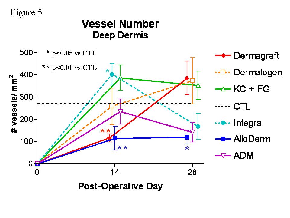

Figure 5.  Graph depicting changes in vessel number (mean±SEM) in the deep dermis at the center of the wound over the study period of 28 days.  The horizontal dashed line represents the vascular counts in normal mouse skin (CTL). Data were analyzed using one-way ANOVA with Tukey’s post tests.


Figure 6.  Graph depicting changes in vessel areas (mean±SEM) in the superficial dermis at the center of the wound over the study period of 28 days.  The dashed horizontal line represents the vessel size in normal mouse skin (CTL). Note that the vessels in wounds treated with Integra are larger than CTL at day 14 (p<0.001) and day 28 (p<0.01).  Data were analyzed using one-way ANOVA with Tukey’s post tests.


   Figure 7.  Graph depicting changes in vessel areas (mean±SEM) in the deep dermis at the center of the wound over the study period of 28 days.  The dashed horizontal line represents the vessel size in normal mouse skin (CTL). Note that the vessels in wounds treated with Integra are larger than CTL at day 14 (p<0.001) and day 28 (p<0.01).  Data were analyzed using one-way ANOVA with Tukey’s post tests.


Figure 8.  Graph depicting percentage of original wound area epithelialized over the study period of 28 days.  Error bars represent SEM.  At day 28, wounds implanted with AlloDerm, Integra, or ADM were approximately 40% reepithelialized, whereas wounds implanted with Dermagraft or Dermalogen were only 10-20% reepithelialized. 
Table 1.  Table of vessel numbers and sizes (mean±SEM) in the total dermis (superficial + deep) for each of the groups at day 28.

FYI.  The content on this blog is copyright protected by the author.  Feel free to read, copy, and disseminate the studies described here, but please indicate the origin of the work by citing this blog URL as an electronic web citation when it is appropriate to recognize and attribute the work of others.

Monday, April 2, 2012

Wound Healing and Neovascularization in Dermal Substitutes

April 3, 2012

This is the first of two interesting wound healing studies done in nude mice implanted with different biological material.  Full thickness skin wounds were created on the dorsum and then different dermal replacements were implanted.  Healing was followed for 28 days and very detailed histological and immunostaining evaluations of the wounds were performed to quantify the healing process.  Most studies of wound healing use simple or highly subjective measures of healing, but one of the main goals of these studies was to use objective criteria to quantify healing and to do so in fine detail.


Healing and Neovascularization of Wounds Implanted with Dermal Substitutes


Healing and Neovascularization of Wounds
Implanted with Dermal Substitutes and Fibrin Glue
in Nude Mice

Performed at the Sumner L. Koch Burn Center, Department of Trauma
John Stroger Jr. Hospital of Cook County 1 &
Department of General Surgery 2
Rush University Medical Center, Chicago, Illinois

Michele M . Loor, MD
Burn Research Fellow (2003-2005)
Department of General Surgery
Rush University Medical Center

Anh-Tuan Truong, MD
Burn Research Fellow (2001-2002)
Metropolitan Group Hospitals Residency in Surgery Program

Barbara A. Latenser, MD 1,2,3
Director, Burn Services

Dorion E. Wiley, MD 1,2
Attending Physician, Department of Trauma, Burn Unit

Robert J. Walter, PhD 1,2
Senior Scientist
Department of Trauma

Address Correspondence to:

Robert J. Walter, PhD
Department of Trauma, Suite 1300
John Stroger Jr. Hospital of Cook County
1900 West Polk Street
Chicago, IL 60612

Phone:  312.864.0578

3 Current address:  University of Iowa Hospitals and Clinics, Department of Surgery, Section of Trauma, Burn, and Critical Care, Iowa City, IA


ABSTRACT

Background:  Dermal substitutes implanted into full-thickness skin wounds reduce wound contraction and improve cosmesis.  These improvements depend upon the development of optimal dermal vascularization.
Methods:  Full-thickness skin wounds were created on the dorsum of nude mice.  A dermal matrix was implanted followed by a mix of fibrin glue (FG) with human keratinocytes (KC).  The following dermal matrices were used: Integra, AlloDerm, ADM, Dermalogen, and Dermagraft.  Wound healing was observed for 4 weeks.  Biopsies were immunostaining for laminin followed by blood vessel quantitation in the superficial and deep dermis in three regions:  wound center, wound margin, and unwounded dermis.
Results: Extensive vascularity was seen at all time points in implanted Dermagraft and Dermalogen.  AlloDerm showed limited vascularity within the first 2 weeks but this normalized by day 28.  ADM and Integra showed rapid but controlled ingrowth of vessels from both the wound base and margins.
Conclusions:  Dermagraft underwent extensive granulation whereas AlloDerm and Dermalogen underwent delayed vascularization.  AlloDerm, Integra, and ADM underwent progressive vessel ingrowth that seemed to be conducive to normal dermal regeneration and modest wound contraction.

Key words:  neovascularization, angiogenesis, wound healing, dermal substitute, nude mouse, Integra, AlloDerm, ADM, Dermagraft, Dermalogen, ACIS


 INTRODUCTION

The treatment of full-thickness wounds in surgery, trauma, and burn poses a significant clinical challenge for several reasons.  An open wound not only provides a portal of entry for microorganisms to invade and proliferate, but it also allows vital fluid and electrolytes to escape. The primary objective in treating an open wound is, therefore, early coverage.  A secondary consideration is optimizing the function and appearance of the healed wound.  Since full-thickness wounds lack a dermis and the dermis does not regenerate, they tend to heal slowly and with significant scarring. Various synthetic collagen-based dermal matrices are now available for use in wounds.1-11  Dermal replacements are intended to provide rapid wound coverage and improve wound healing.  Once implanted, these replacements either become incorporated into the wound or stimulate tissue growth. However, the efficacy of these materials in the treatment of full-thickness wounds has not been studied carefully and compared.

In most cases, placement of a dermal matrix is later followed by additional surgery for split-thickness skin grafting in order to achieve definitive wound closure. The use of cultured autologous keratinocytes has been studied extensively as an alternative to autografting, particularly in patients with greater than 40% total body surface area wounds who have limited donor site availability.12-19 Various methods of keratinocyte (KC) delivery to wounds are available including combination with a fibrin sealant.  Several studies have suggested the utility of fibrin glue (FG) in this setting, with evidence to suggest enhanced reepithelialization and basement membrane formation.20-24 Cultured KCs can also be introduced into wounds via a spray apparatus.25  Preliminary studies in which KCs were suspended in fibrin glue and sprayed onto wounds indicate that this permits acceptable cell survival and proliferation.26-29 Other potential advantages of the use of fibrin glue in wounds are improved hemostasis and protection from infection.30 A KC spray apparatus has been developed in our lab for use following suspension of the KCs in the thrombin component of fibrin sealant (Tisseel®; Baxter, Deerfield, IL).  It has tested both in vitro and in vivo, and the viability and proliferative potential of the KCs immediately and 24-48 hours after spraying are affected very little.

Neovascularization is a key step in wound healing, as new vessels are necessary to support the newly formed tissue.  It is a complex process that is dependent upon appropriate interactions between the extracellular matrix, the migrating endothelial cells, and a number of growth factors.31  Dermal substitutes composed of native (undenatured), allogenic extracellular matrix such as acellular dermal matrix (ADM) and AlloDerm become readily vascularized, complement healing, and reduce contractive scarring in a variety of wound types.  We hypothesized that the mode and rate of neovascularization during the process of wound healing in the presence of ADM or AlloDerm may be an important determinant in final wound resolution.  The process of vascularization may be significantly altered with the use of other materials such as those containing synthetic, xenogenic, or denatured substances including Integra, Dermalogen, and Dermagraft, thereby negatively affecting wound resolution.

To evaluate this, dermal substitutes in conjunction with FG and KC were introduced into full-thickness wounds on the dorsum of nude mice.  The progress of wound healing and particularly dermal neovascularization was evaluated by objective criteria over a period of four weeks.  Paraffin-embedded sections from weekly biopsies were immunostained for laminin, an antigen found in the endothelial basement membrane, to assess and compare vascularization in these healing wounds.


METHODS AND MATERIALS

      The following materials were used:
§         Tisseel® (Baxter Health, Deerfield, IL) is a two component system in which fibrinogen, calcium, thrombin, and a protease inhibitor are combined and dispensed onto a wound or other surface to form a fibrin clot. 
§         Integra® (Ethicon, Somerville, NJ) is a bilayer artificial skin composed of a “dermal” layer of bovine collagen gel cross-linked with shark chondroitin-6-sulfate and an “epidermal” layer of polysiloxane polymer (which was removed for this study). 
§         AlloDerm® (Lifecell Corp., Branchburg, NJ) is a collagen matrix derived from human skin that is treated to remove most of the cellular components.
§          Acellular Dermal Matrix (ADM) is a dermal collagen matrix derived from human skin that is treated to remove all cellular components.11, 31Dermalogen® (Collagenesis Corp, Beverly, MA) is a powdered human dermal matrix that has been treated to remove some cellular components and contains collagens, fibronectin, and elastin.  This matrix material is supplied as a 3.5% suspension in phosphate buffer. 
§         Dermagraft® (Advanced Tissue Sciences) is comprised of a woven bioabsorbable polymer on and in which human dermal fibroblasts are grown and then devitalized.

Animals and Surgery

NIH homozygous male nude mice, 4 weeks of age (Taconic, Germantown, NY) were used.  This model permits the implantation of xenogenic materials, such as those present in these dermal substitutes (e.g., bovine collagen, shark chondroitin sulfate, human collagens, human fibroblasts and KCs).  The disadvantage of this model is that the effect on neovascularization of immune reactivity against such xenogenic materials that might be evinced in humans will be masked or absent.

All surgical interventions and experiments were performed in the John H. Stroger, Jr. Hospital of Cook County Animal Care Facility using protocols approved by the IACUC.  Preoperative antibiotics (Kanamycin 25U/kg IM x 1 dose) were administered to the animals and ketamine/ xylazine was used for anesthesia.  Under aseptic conditions, 2 cm x 2 cm full-thickness wounds were excised down to the muscle fascia, removing the panniculus carnosus.  The groups used in this experiment were as follows: 1) Integra, 2) AlloDerm, 3) ADM, 4) Dermalogen, 5) Dermagraft, 6) KCs + FG only, and 7) FG only.  Animals in groups 1-6 received human KCs sprayed onto the wound surface in combination with FG.  Each group was comprised of at least 6 mice, three of which underwent biopsies at days 7, 14, 21 and 28.  Three more mice were treated as in groups 1-6 except that FG was administered without KCs.  In the groups of mice that received dermal substitutes, the substitute was cut to size and sutured to place in the wound using 4-0 nylon sutures.  In the Dermalogen group, 2 cc of a suspension was placed in the wound.  In the groups of mice to receive KCs, KCs at a concentration of 104 per cc in Tisseel (0.5 cc) were sprayed on the dermal matrix.  All defects were covered with a semi-permeable adhesive film (Op-Site, Smith & Nephew, Largo, FL), Xeroform (Sherwood Medical, St. Louis, MO), dry cotton gauze (Adaptic, Johnson & Johnson, New Brunswick, NJ), and finally with a fine stainless steel mesh fixed to the animals’ back with skin sutures.  This last was used to prevent the wounds from being disturbed by chewing or scratching.  Dressings were inspected daily.  At the completion of the study period (4 weeks), all animals were euthanized.  The harvested biopsies were fixed in 10% buffered formalin.  Specimens were paraffin embedded and sections stained with H&E or immunostained. 

Preparation of ADM

Thawed cadaver skin was treated with 2.5 units/ml Dispase II (Boehringer Mannheim, Indianapolis, IN) in PBS containing 0.2 mM CaCl2 at 4EC for 24 hours to remove the epidermis and other cellular components from the dermal matrix.  Subsequently, the dermal matrix was incubated in buffered 0.5% Triton X-100 (U. S. Biochemical Corp., Cleveland, OH) for 24 hours at room temperature with continuous shaking.  ADM was then extensively washed with PBS and stored in PBS at 4EC until use 11,31.

Human KC Culture and Preparation for Spraying

Human KCs, Epilife culture medium, supplements, and transfer solutions (trypsin, trypsin neutralizer) were obtained from Cascade Biologics (Portland, OR).  KCs arrived tested and warranted to be free of HIV, hepatitis B and C, mycoplasma, bacteria, yeast and other fungi.  Cells were grown in 75 cm2 flasks from expanded frozen stocks stored after passage 2.  After 7-10 days of proliferation and growth, flasks containing 50-80% confluent KCs were washed, trypsinized briefly to release cells from the substrate, trypsin neutralized, and the suspension centrifuged at 20xg for 5 min and 4ºC.  The supernatant was discarded and the cells resuspended in fresh Epilife medium. Cells were mixed with the reconstituted thrombin/calcium component of the fibrin sealant kit at a 1:1 dilution.  The fibrinogen component was also reconstituted and diluted 1:1 with Epilife.  Both components were stored at 4ºC until sprayed onto wounds in mice as indicated above.   Viability of the cells following mixing with the thrombin component of Tisseel and after spraying using spray apparatus was confirmed using trypan blue staining.

Immunostaining

Paraffin sections were deparaffinized and then antigen retrieval was performed by incubating specimens in pepsin (1mg/ ml, in 0.01N HCl) at 37ºC for 2 hours.  Following antigen retrieval, blocking of nonspecific binding was accomplished by incubation in a solution of 1% bovine serum albumin in Tris buffered saline, pH 7.9 for 20 min.  Sections were then incubated in rabbit anti-mouse laminin IgG (Sigma, St. Louis, MO) at a 1:10 dilution for 2 hr at room temperature followed by HRP-conjugated goat anti-rabbit IgG secondary antibody (Cappel, Irvine, CA) at a dilution of 1:100 for 1 hr at room temperature.  Reaction product was generated using a Vector DAB/peroxide Developing Kit (Vector, Burlingame, VT) according to the manufacturer’s specifications with a 10 min developing time.  Specimens were counterstained with Vector Hematoxylin QS (Vector, Burlingame, VT) for one minute.  Normal skin from previously unwounded mice was also immunostained for laminin as above to serve as a control.  Laminin staining identified the basement membrane of epidermis, nerves, muscle, and vessels.  Attention was focused on the dermis, within which stained structures with a clear lumen were counted as blood vessels.  These were readily distinguished from the other positively stained structures by their location and shape.  Immunostaining for endoglin (CD105) and CD34 was performed but the stain resulting was either nonspecific or too weak to be of use here.

Data Analysis

Wound characteristics were measured grossly and histologically.  Gross observations were made at days 7, 14, 21, and 28 post-surgery.  Wound contraction and degree of epithelialization were measured using UTHSCSA ImageTool software in conjunction with digital photographs of wounds.  Histological characteristics determined by H&E staining included: the presence and thickness of a stratified epithelial layer, the persistence of the implanted dermal matrix, and the degree of inflammation.  The degree of vascularity was determined by two methods.  Manual vessel counts were carried out on the sections using an eyepiece reticle.  The size of this reticle grid at the final magnification used (400X) was 250 x 250 μm.  Numbers of blood vessels were determined by counting vessels in the superficial dermis, i.e., with one side of the reticle on the epidermal basement membrane and the counts performed in a 250 μm square of dermis underlying the basement membrane or in the deep dermis, i.e., with one side of the reticle positioned on the hypodermis and the counts performed in 250 μm square of dermis directly overlying the hypodermis. 

Three different zones of each wound biopsy were evaluated in this way: the wound center (WC), wound margin (WM) and unwounded normal dermis peripheral to the wound margin (NP).  Slides were scanned visually at 40X to find wholly intact sections showing all 3 regions of interest (WM, WC, and NP) and then viewed at 400X magnification for vessel counts.  Within each zone, two or more immediately adjacent regions were counted and averaged.  Placement of the reticle was standardized by defining: the WC as the region equidistant from each wound margin; the WM as the area directly adjacent to unwounded tissue; and the NP as the area with normal skin structure and appendages at least 500 µm away from the WM.  Additionally, some vessel counts were performed on laminin immunostained tissue sections using the ChromaVision (San Juan Capistrano, CA) Automated Cellular Imaging System (ACIS) in conjunction with microvascular density (MVD) software.  Image analysis was performed in each of these zones with one set of six counts obtained for each region from which the microvascular density (#vessels/ mm2) was calculated.  Wounds that demonstrated excessive blood vessel proliferation (more than 3 times the level seen in unwounded mouse skin) were considered granulation tissue.  Data from days 14 and 28 were analyzed with one-way ANOVA and Tukey post-tests. 

RESULTS
Gross wound observations
 Each of the dermal substitutes except Dermalogen reduced the amount of wound contraction as compared to wounds that received no dermal substitute and sprayed KCs + FG (figure 1).  Thirty-five to 45% wound contraction was observed in wounds implanted with AlloDerm, Dermagraft, Integra, or ADM at 28 days post-operatively.  Greater contraction (60%) was observed in the Dermalogen and KCs + FG groups (figures 2, 3).
H&E staining
 Histologically, KCs sprayed into the wounds remained within the FG and sometimes formed a monolayer by day 14.  AlloDerm, ADM, and Integra became vascularized and infiltrated with fibroblasts within 7-14 days.  AlloDerm and ADM became integrated into the wound by 28 days (figure 4) whereas Integra underwent extensive breakdown coincident with the appearance of numerous multinucleated giant cells.  Dermagraft formed a covering over the wound that did not dissolve or remodel and did not promote or permit the development of a neodermis.  Dermalogen was extensively resorbed post-operatively and remained highly disorganized such that poor healing resulted.  In wounds lacking any dermal substitute, KCs tended to migrate toward the wound base but did not form a monolayer.  The lack of a dermis resulted in extensive contraction of these wounds.

Laminin Immunostaining and Vessel Counts

Vessel counts were performed on immunostained sections from biopsies taken on days 14 and 28 (figures 5 and 6).  Six different regions were scored for each section: wound center superficial (WCS) and deep (WCD), wound margin superficial (WMS) and deep (WMD), normal peripheral dermis superficial (NPS) and deep (NPD) (Figure 7).  The primary control in this study was normal skin from unwounded animals (CT) which was analyzed for vessel counts in the superficial and deep regions.  For each of the groups, the number of vessels in the normal peripheral (NP) tissue flanking the wound was an additional internal control.  These two controls (CT and NP) were compared (figure 8) for each of the dermal substitute groups and were found to be not significantly different (ANOVA, p=0.23).  A group of animals treated with allogenic or autogenic skin grafts was included.  Gross wound healing was very good in this group but immunostaining labeled pre-existing and newly formed vessels indistinguishably.

On day 14, the Integra, Dermagraft, and Dermalogen groups showed statistically significant differences in the number of vessels in the WCS and WCD regions compared to CT skin (figure 9; ANOVA, p < 0.01).  Wounds treated with AlloDerm (for WCD) or ADM (for WCS) had significantly fewer vessels in the WC than CT (Tukey test, p< 0.05).  For Dermalogen and Dermagraft, there were increased numbers of vessels in the wound margin (p<0.01).  By day 28 (figure 10), the vessel counts for each of the groups had normalized, with no statistically significant differences between any of the groups and CT except for AlloDerm, which was hypervascular in the WCS (p<0.01), and KC + FG and Dermagraft which were hypervascular in the WMS (p<0.01).

In the superficial dermis, the number of vessels in most groups approached the level of CT at day 28 (figure 11).  However, the vascularity in the superficial dermis with AlloDerm between days 21 and 28 underwent a striking increase resulting in hypervascularity (p < 0.01).  Dermalogen, ADM, and Integra showed limited vascularity in the superficial dermis on day 14 (p<0.01, p<0.05, and p<0.01, respectively) until day 28, when the number of vessels was similar to normal dermis.  In contrast, hypervascularity was seen in the superficial dermis for the Dermagraft group beginning at day 7 (data not shown) with a peak at day 14 (p < 0.05) and normalization by day 28.  Vascularity in wounds treated with FG only was similar to CT from day 7 to day 28.

In the deep dermis, the patterns of vessel ingrowth were similar to those described above for the superficial dermis (figure 12). At day 14, there was a statistically significant elevation in vessel number for Dermagraft and Integra (p<0.01), with very little vascularity in the AlloDerm (p<0.05) or Dermalogen (p<0.01) groups.  All of the wounds approached normal levels of vascularity by day 28, with no statistically significant differences from CT.

Automated Vessel Counts

            Image analysis was used to evaluate the impact of KCs on vascularization of wounds treated with dermal substitutes and FG.  At day 28 in the wound center, significantly more blood vessels were present when KCs were included (p=0.027, paired t-test) (figure 13).  In addition, the average area per blood vessel (µm2/ vessel) was calculated for these groups.  In CT skin, the average vessel size was 300 µm2.  In the groups without KCs, the average blood vessel size was 360 µm2.  In the groups sprayed with KCs + FG, the average blood vessel size was equal to that in normal CT mouse skin at 300 µm2. 


DISCUSSION

Angiogenesis is an integral part of normal wound healing.  Blood vessels deliver oxygen, nutrients and inflammatory cells into the wound, and also remove necrotic tissue from the area.  Appropriate angiogenesis is a complex process that includes endothelial cell division, selective degradation of vascular basement membrane and of surrounding extracellular matrix, and endothelial cell migration.31 Each step requires an appropriate balance between activators or growth factors and inhibitors. 32  In addition, the level of organization of the extracellular matrix plays a key role in the regulation of neovascularization, in that it provides support for migrating endothelial cells and acts as a reservoir for growth factors.33  Ideally, full-thickness wounds undergo an initial phase of vigorous angiogenesis that is later followed by vessel regression, such that the final pattern of vascularization is similar to that of normal skin.32

            However, no studies have been published comparing wound healing and particularly angiogenesis using the different commercially-available dermal substitutes.  Most studies compare one substitute to split-thickness skin grafting and most use subjective criteria for evaluating the results.15 Thus, it is difficult to objectively determine which dermal substitutes are most useful in the treatment of full-thickness wounds.  Within this context, there are few studies which have examined the process of neovascularization in wounds implanted with dermal substitutes.  We hypothesized that wounds implanted with dermal substitutes composed of undenatured, allogenic extracellular matrix, such as ADM and AlloDerm, wound be readily incorporated and have a final level of vascularization more similar to normal skin than dermal substitutes composed of synthetic, xenogenic, or denatured substances, such as Integra, Dermalogen, and Dermagraft.  To a great extent, this hypothesis was confirmed by the data presented here.

Grossly, wounds implanted with ADM, AlloDerm, or Integra demonstrated less wound contraction (about 40% by day 28) and better cosmetic results than did Dermagraft, Dermalogen, or KC + FG only.  Our findings indicate that of the dermal substitutes with native compositions, ADM implants achieved a normal level of vascularization gradually over the 28 day period.  On the other hand, AlloDerm which has a composition similar to ADM, underwent a gradual increase in the number of vessels but was hypervascular in the superficial dermis at day 28.  For the synthetic dermal substitutes, vascularization proceeded gradually and approached normal at day 28, with the exception of Dermagraft which was highly vascularized at days 7 (data not shown) and 14.

The results for Integra were noteworthy in that despite its denatured, xenogenic composition, a controlled pattern of vessel ingrowth conducive to improved wound healing was seen.  However, the collagen-GAG matrix of Integra has been specifically designed to have the necessary pore size essential to ensure adequate microvascularization of the neodermis.34-37   Wounds in the Dermagraft group demonstrated extensive granulation which may to some extent be attributable to the presence of fibroblasts and therefore vascular endothelial growth factor (VEGF) in the matrix.  Similarly, when fibroblasts are added to other dermal substitutes, such as de-epidermized dermis, enhanced wound vascularization has been shown.38  However, in our study increased vascularization during the healing process does not appear to improve wound healing.  We also observed significantly increased amounts of vascularization in wounds treated with KCs + FG in comparison to wounds which received FG alone.  KCs are known to produce VEGF which is a potent inducer of angiogenesis.31  In wounds treated with KCs that overexpress VEGF, decreased wound contraction, improved tissue development, and increased vascular density are seen in the dermis.39  Nonetheless, in the present study the final outcome in wounds treated with only KCs + FG or FG alone was clearly scarring with extensive wound contraction.  Thus, the supranormal increase in vascular density present during the course of healing correlated with increased wound contraction.

Overall, there seems to be an optimal level of vascularization in healing wounds, where either increased or decreased levels lead to suboptimal wound healing.  A certain level of vascularity of any dermal substitute is required for the subsequent take of STSG, CEA, or other KC preparations.  The observed differences between the healing seen with the dermal substitutes studied here will depend on a number of factors including differences in levels of tissue oxygenation, in the growth factor milieu, and in the structure and composition of the extracellular matrices.  Dermagraft underwent extensive early granulation, whereas AlloDerm and Dermalogen underwent delayed vascularization.  AlloDerm, Integra, and ADM underwent progressive vessel ingrowth that seemed to be conducive to normal dermal regeneration, reepithelialization, and limited wound contraction.  These results indicate that the rate and final extent of vascularization are important determinants of the efficacy of dermal substitution for treating full-thickness wounds.  By evaluating wound contracture, epithelialization, and angiogenesis in this nude mouse model, the value of novel biomaterials as dermal substitutes may be predicted.

In the treatment of chronic wounds, stimulation of angiogenesis effectively promotes wound closure in patients with diabetes or peripheral vascular disease.  However, in the treatment of acute full-thickness wounds, hypervascularity or granulation must be limited to reduce contraction and scarring.  As seen here, AlloDerm, ADM, and Integra demonstrated the desired pattern of vascularization whereas other materials tested tended to become hypervascularized.  Thus, we may expect that the use of AlloDerm, ADM, or Integra should lead to improved healing of full-thickness wounds in patients. 


ACKNOWLEDGMENTS

The authors would like to thank Paolo Gattuso, MD for giving them access to the ChromaVision system and Christopher Valadez and ChromaVision technical support for their assistance in using the system.


 REFERENCES

1.        Burke JF, Yannas IV, Quinby WC, Bondoc CC, Jung WK. Successful use of a physiologically acceptable artificial skin in the treatment of extensive burn injury. Annals Surg. 1981; 194: 413-427.

2.        Hansbrough JF, Boyce ST, Cooper ML, Foreman T. Burn wound closure with cultured autologous keratinocytes and fibroblasts attached to a collagen-glycosaminoglycan substrate. JAMA. 1989; 262: 2125-2130.

3.        Boyce ST, Greenhalgh DG, Kagan RJ, et al. Skin anatomy and antigen expression after burn wound closure with composite grafts of cultured skin cells and biopolymers. Plast Reconstr Surg. 1993; 91: 632-64.

4.        Bell E, Ehrlich HP, Buttle DJ, Nakatsuji T. Living tissue formed in vitro and accepted as skin-equivalent tissue of full-thickness. Science. 1981; 211:1052-1054.

5.        Heimbach D, Luterman A, Burke J, et al. Artificial dermis for major burns. Annals Surg. 1988; 208: 313-319.

6.        Matsuda K, Suzuki S, Isshiki K, et al. A bilayer "artificial skin" capable of sustained release of an antibiotic. Brit J Plast Surg. 1991; 44: 142-146.

7.        Cooper ML, Hansbrough JF. Use of a composite skin graft composed of cultured human keratinocytes and fibroblasts and a collagen-GAG matrix to cover full-thickness wounds on athymic mice. Surgery. 1991; 109: 198-207.

8.        Hansbrough JF, Morgan J, Greenleaf G. Evaluation of Graftskin composite grafts on full-thickness wounds on athymic mice. J Burn Care Rehabil. 1994; 15: 346-353.

9.        Hansbrough JF, Morgan J, Greenleaf G, Bartel R. Composite grafts of human keratinocytes grown on a polyglactin mesh-cultured fibroblast dermal substitute function as a bilayer skin replacement in full-thickness wounds on athymic mice. J Burn Care Rehabil. 1993; 14: 485-494.

10.     Matouskova E, Vogtova D, Konigova R. A recombined skin composed of human keratinocytes cultured on cell-free pig dermis. Burns.  1993; 19: 118-123.
11.     Takami Y, Matsuda T, Yoshitake M, Hanumadass M, Walter RJ. Dispase/detergent treated dermal matrix as a dermal substitute. Burns. 1996; 22: 182-190.
12.     Teepe RGC, Kreis RW, Korbrugge EJ, et al. The use of cultured autologous epidermis in the treatment of extensive burn wounds. J Trauma. 1990; 30: 269-275.
13.     Nanchahal J, Ward CM. New grafts for old?  A review of alternatives to autologous skin. Brit J Plast Surg. 1992;  45: 354-363.
14.     Shakespeare P. Burn wound healing and skin substitutes. Burns. 2001;  27: 517-522.
15.     Kearney JN. Clinical evaluation of skin substitutes. Burns. 2001;  27: 545-551.
16.     Balasubramani M, Kumar TR, Babu M. Skin substitutes: a review. Burns.  2001;  27: 534-544.
17.     Boyce ST. Design principles for composition and performance of cultured skin substitutes. Burns. 2001;  27: 523-533.
18.     Rue LW, Cioffi WG, McManus WF, Pruitt BA. Wound closure and outcome in extensively burned patients treated with cultured autologous keratinocytes.  J Trauma.  1993; 34: 662-667.
19.     Gallico GG, O’Connor NE, Compton CC, Kehinde O, Green H.  Permanent coverage of large burn wounds with autologous cultured human epithelium.  NEJM. 1984; 311: 448-451.
20.     Cohen M, Bahoric A, Clarke HM. Aerosolization of epidermal cells with fibrin glue for the epithelialization of porcine wounds with unfavorable topography. Plast Reconstr Surg. 2001; 107: 1208-1215.
21.     Currie LJ, Martin R, Sharpe JR, James SE.  A comparison of keratinocyte cell sprays with and without fibrin glue.  Burns. 2003; 29: 677-685.
22.     Horch RE, Bannasch H, Kopp J, Andree C, Stark GB.  Single-cell suspensions of cultured human keratinocytes in fibrin glue reconstitute the epidermis.  Cell Transplant. 1998; 7: 309-317.
23.     Hunyadi J, Farkas B, Bertenyi C, Olah J, Dobozy A.  Keratinocyte grafting: a new means of transplantation for full-thickness wounds.  J Dermatol Surg Onc. 1988; 14: 75-78.
24.     Ronfard V, Rives J-M, Neveux Y, Carsin H, Barrandon Y.  Long-term regeneration of human epidermis on third degree burns transplanted with autologous cultured epithelium grown on a fibrin matrix.  Transplantation.  2000; 70: 1588-1598.
25.     Fraulin FOG, Bahoric DVM, Harrop AR, Hiruki T, Clarke HM.  Autotransplantation of epithelial cells in the pig via an aerosol vehicle.  J Burn Care Rehabil 1998; 19: 337-345.
26.     Navarro FA, Stoner ML, Lee HB, Park CS, Wood FM, Orgill DP. Melanocyte repopulation in full-thickness wounds using a cell spray apparatus. J Burn Care Rehabil. 2001; 22: 41-46.
27.     Navarro FA, Stoner ML, Park CS, Huertas JC, Lee HB, Wood FM, Orgill DP. Sprayed keratinocyte suspensions accelerate epidermal coverage in a porcine microwound model. J Burn Care Rehabil. 2000;. 21: 513-518.
28.     Jiao XY, Kopp J, Tanczos E, Voigt M, Stark GB.  Cultured keratinocytes suspended in fibrin glue to cover full-thickness wounds on athymic nude mice: comparison of two brands of fibrin glue.  Eur J Plast Surg. 1988; 21: 72-76.
29.     Chester DL, Balderson DS, Papini RPG.  A review of keratinocyte delivery to the wound bed.  J Burn Care Rehabil. 2004; 25: 266-275.
30.     Currie LJ, Sharpe JR, Martin R.  The use of fibrin glue in skin grafts and tissue-engineered skin replacements: a review.  Plast Reconstr Surg. 2001; 108: 1713-1726.
31.     Dvorak HF, Brown LF, Detmar M, Dvorak AM.  Vascular permeability factor/vascular endothelial growth factor, microvascular hyperpermeability, and angiogenesis.  Am J Pathol. 1995; 146: 1029-1039.
32.     Lingen MW.  Role of leukocytes and endothelial cells in the development of angiogenesis in inflammation and wound healing.  Arch Pathol Lab Med. 2001; 125: 67-71.
33.     Li J, Zhang Y-P, Kirsner RS.  Angiogenesis in wound repair: angiogenic growth factors and the extracellular matrix.  Microsc Res Tech.  2003; 60: 107-114.
34.     Burke JF.  Observations on the development of an artificial skin: presidential address, 1982 American Burn Association Meeting. J Trauma. 1983; 23: 543-551.
35.     Moiemen NS, Staiano JJ, Ojeh, NO, Thway Y, Frame JD. Reconstructive surgery with a dermal regeneration template: clinical and histologic study.  Plast Reconstr Surg. 2001; 108: 93-103.

36.     Stern R, McPherson M, Longaker MT.  Histologic study of artificial skin used in the treatment of full-thickness thermal injury. J Burn Care Rehabil. 1990; 11: 7-13.

37.     Sheridan RL, Hegarty M, Tompkins RG, Burke JF.  Artificial skin in massive burns-results to ten years.  Eur J Plast Surg.  1994; 17: 91-93.

38.     Erdag G, Sheridan RL. Fibroblasts improve performance of cultured composite skin substitutes on athymic mice.  Burns. 2004; 30: 322-328.

39.     Supp DM, Boyce ST.  Overexpression of vascular endothelial growth factor accelerates early vascularization and improves healing of genetically modified cultured skin substitutes.  J Burn Care Rehabil. 2002; 23: 10-20.


FIGURES


Figure 1

Wound area over the 28 day study period for each of the dermal substitutes.  Values are normalized against the size of each wound on the day of surgery.  Dashed horizontal line represents starting wound area in normal unwounded mouse skin.

Figure 2

Gross appearance of wounds on the day of surgery shows the structural differences between the dermal substitutes.  Full-thickness wounds (2 x 2 cm) were created, followed by implantation of the dermal substitute and sprayed FG and KCs.  Nylon sutures were placed at all four corners of the wound to mark the original wound size.
Figure 3

Gross appearance of wounds on post-operative day 28 shows the healed wounds and the degree of contraction.  Sutures mark the corners of the original wound.  Note the extensive contraction of wounds treated with KC + FG only and Dermalogen in comparison to the minimal contraction observed with use of Integra, AlloDerm, and ADM. Dermagraft-implanted wounds showed poor epithelialization and incorporation into the wound with contraction limited only as long as the implant was retained.

Figure 4

H&E stained paraffin cross-sections of day 28 biopsies for each of the dermal substitute groups.  The Integra specimen shows scattered KCs on the surface of the FG which is overlying the Integra and numerous small islands of residual gel within the developing neodermis (arrows).  The Dermagraft specimen shows synthetic fibers that have been incorporated into the wound (arrows).  The AlloDerm and ADM specimens are epithelialized and show a structured dermal matrix populated with fibroblasts, blood vessels, and other connective tissue components.  Magnification bar =  200 μm

Figure 5

Cross-sections of wound biopsies immunostained for laminin showing the wound center for Integra and AlloDerm on day 14.  Vessels can be clearly identified (arrows) within the deep dermis for Integra.  The superficial region of the Integra implant and the entire AlloDerm implant are evident and show few vessels.  Magnification bars = 100 μm

Figure 6

Cross-sections of wound biopsies immunostained for laminin showing the wound center for Integra and AlloDerm on day 28.  AlloDerm has numerous vessels scattered throughout the dermis as does Integra.  Note the numerous large and small cavities in the Integra implant.  In life, these cavities held the collagen-chondroitin sulfate colloid that comprises Integra.  Magnification bars = 100 μm

Figure 7

Laminin-stained paraffin cross-section of Integra on day 28 illustrating the different regions in which vessel counts were performed.  Three different zones were identified: Normal peripheral (NP) tissue, wound margin (WM), and wound center (WC).  Within each zone, counts were performed in the superficial regions (S), the area directly below the epidermal basement membrane and in the deep regions (D).  Note that the normal peripheral zones that were actually counted were further away from the wound margin than shown (>300 μm).  In total, six different regions (NPS, NPD, WMS, WMD, WCS, and WCD) were analyzed for each section.  Magnification bar = 150 μm

Figure 8

Stacked bar graph showing blood vessel counts in superficial (sup) and deep regions of normal peripheral tissue for each dermal substitute on days 14 and 28 post-surgery.  The dotted line represents the total number of blood vessels (i.e., superficial + deep) seen in control normal mouse skin (CT).  Error bars represent standard error of the mean.  One-way ANOVA did not show any significant differences between groups and CT, p = 0.238 

Figure 9

Stacked bar graph shows blood vessel counts (mean ± SEM) in the superficial (sup) and deep regions of the wound center and wound margin on day 14 post-surgery for each dermal substitute.  The dotted line represents the total number of blood vessels (i.e., superficial + deep) in control normal mouse skin (CT).  Data were analyzed using one-way ANOVA with Tukey post-tests.

Figure 10

Stacked bar graph shows blood vessel counts (mean ± SEM) in the superficial (sup) and deep regions of the wound center and wound margin on day 28 post-surgery for each dermal substitute.  The dotted line represents the total number of blood vessels (i.e., superficial + deep) in control normal mouse skin (CT).  Data were analyzed using one-way ANOVA with Tukey post-tests.

Figure 11

Graph depicting changes in vessel numbers (mean ± SEM) in the superficial dermis at the center of the wound during the 28-day study period.  The dashed horizontal line represents the vascular counts in normal mouse skin (CT).  Note the great elevation in vessel numbers for Dermagraft implants especially at day 14.  By day 28, vascular counts were similar to CT for most of the groups.  Data were analyzed using one-way ANOVA with Tukey post-tests.
**p < 0.05 for Dermalogen, Integra, or ADM vs CT
  *p < 0.01 for AlloDerm on day 28 and for Dermagraft on day 14 vs CT

Figure 12

Graph depicting changes in vessel numbers (mean ± SEM) in the deep dermis at the center of the wound during the 28-day study period.  The horizontal dashed line represents the vascular counts in normal mouse skin (CT).  Note the great elevation in counts for Dermagraft implants especially at day 14.  By day 28, vascular counts were similar to CT for all groups.  Data were analyzed using one-way ANOVA with Tukey post-tests.
**p < 0.05 for AlloDerm vs CT
  *p < 0.01 for Dermagraft, Integra, or Dermalogen vs CT

Figure 13

Vessel counts from ChromaVision ACIS system at the wound center for wounds treated with dermal substitute + FG + KC versus wounds treated with dermal substitute + FG alone on day 28.  The horizontal dashed line represents the vascular counts in normal mouse skin.  Overall, there are significantly (p=0.027, paired t-test) more blood vessels seen when KCs are present.

 FYI.  The content on this blog is copyright protected by the author.  Feel free to read, copy, and disseminate the studies described here, but please indicate the origin of the work by citing this blog URL as an electronic web citation when it is appropriate to recognize and attribute the work of others.