Showing posts with label neutrophils. Show all posts

Tuesday, May 1, 2012

Mechanism of Cancer Chemotaxis Defect

May 2, 2012

When we were studying the chemotaxis defect known to exist in leukocytes from cancer patients, we looked at formylpeptide receptor dynamics in considerable detail.  We examined receptor binding, down-regulation, recovery, and receptor-mediated pinocytosis using rabbit peritoneal PMN, human peripheral PMN, and human peripheral monocytes.  The cells were either obtained directly from cancer patients or were from normal subjects.  Cells from normal subjects were pretreated with serum that had been obtained from cancer or normal patients.

The full paper and link to the PDF are shown below.

 Mechanism of Cancer Chemotaxis Defect


Mechanism of the Cancer-Related Leukocyte Chemotaxis Defect: 

Formylpeptide Receptor Modulation and Pinocytosis


Amelia H. Janeczek, PhD1,3, Pierson J. Van Alten, PhD1, Hernan M. Reyes, MD2,

and Robert J. Walter, PhD2


1  Department of Anatomy and Cell Biology, University of Illinois at Chicago, Chicago, IL  60612

2  Department of Surgery, Cook County Hospital, Hektoen Institute for Medical Research, Chicago, IL  60612

3  Present address:  Department of Biochemistry, School of Medicine, Boston University, Boston, MA  02118-2394


Address all correspondence to:

Robert J. Walter, PhD
Department of Surgery, Room 905
Hektoen Institute for Medical Research
625 South Wood Street
Chicago, IL  60612


Telephone:        (312) 633-7237/8717           FAX:       (312) 738-3102

Running Title:  Mechanism of Cancer Chemotaxis Defect

Keywords:  formylpeptide, receptors, cancer, chemotaxis, endocytosis


SUMMARY

Monocyte chemotaxis is severely depressed in patients with advanced tumors but the cellular basis for this chemotactic defect is not known.  Pretreatment of normal human leukocytes or rabbit peritoneal neutrophils (PMN) with serum from cancer (CA) patients inhibits both monocyte and PMN chemotaxis as compared to leukocytes pretreated with serum from healthy (CT) blood donors.  Using purified fresh CA patient leukocytes or CA serum-treated normal leukocytes, formylpeptide receptor binding and modulation were quantified using radiolabeled formylmethionyl-leucyl-phenylalanine (3H-FMLP).  The cell surface binding of 3H-FMLP at 4°C was significantly reduced in CA serum pretreated rabbit peritoneal PMN, but not in CA serum pretreated human peripheral blood PMN or mononuclear leukocytes as compared to CT serum pretreated cells.  However, in purified mononuclear leukocytes isolated directly from tumor patients, formylpeptide binding was significantly reduced as compared to those of normal subjects.  PMN from tumor patients exhibited no significant difference in this regard as compared to PMN from control subjects.  The time course and dose response curves observed during formylpeptide receptor down-regulation were similar for CT and CA serum pretreated cells.  Subsequent to down-regulation, the recovery of cell surface formylpeptide binding at 37°C was similar in its rate and extent in CT and CA serum pre­treated cells.  However, significant reductions in 3H-FMLP uptake at 37°C were seen in tumor patient PMN, in CA serum pretreated rabbit PMN, and in CA serum pretreated human PMN and mononuclear leukocytes as compared to controls.  Such reductions in initial formylpeptide cell surface binding and in formylpeptide endocytosis may contribute directly to the cancer-associated depression of leukocyte chemotaxis.


INTRODUCTION

      Monocyte and macrophage chemotaxis is severely impaired in patients with advanced tumors, but neutrophil (PMN) locomotion is unaffected (1,2).  This defect in monocyte chemotaxis may have life-threatening consequences since host leukocytes may be unable to adequately repress bacterial and fungal infections or tumor growth.  Implantation of tumor cells or injection of tumor sonicates into mice results in reduced accumulation of macrophages in response to inflammatory stimuli, as well as decreased resistance to bacterial infec­tion (3,4).  Similarly, treatment of normal leukocytes with serum from patients with advanced tumors, conditioned media from tumor cell lines, as­cites fluid, plasma, or urine from tumor-bearing mice suppresses monocyte (and often PMN) polarization and chemotaxis (2,5‑9).  This chemo­taxis defect is evident in animals and patients bearing any of a large number of tumors and is seen in response to several quite different classes of che­moattractants (for review see 6,10,11).  Many studies have shown that monocyte chemo­taxis returns to normal after the surgical removal of tumor or after treatment with chemotherapy or radiotherapy (12,13).  Taken together, this suggests that tumors may be producing or causing the host to produce an inhibitor of leukocyte chemotaxis (7,14). 


A soluble inhibitor of monocyte chemotaxis, rendered inactive by treat­ment with antibody directed against the retroviral envelope protein p15E, has been found in tumor patient effusions (15,16) and in serum from patients with head and neck cancer (CA) (17).  This inhibitor or its synthetic ana­logues may cause decreased monocyte polarization in response to chemoattract­ant (8,16), alterations in formylpeptide receptor expression (18,19), suppression of the respiratory burst (20), and inhibition of protein kinase C-related cell functions (21).  It was hypothesized in the present study that the chemotactic defect in tumor patient monocytes or in CA serum-treated leukocytes resulted from alterations in reception or transduction of the signal for chemotaxis.  To examine this possibility, formylpeptide chemoattractant receptor binding was examined in leuko­cytes isolated from tumor patients, in CT or CA serum pretreated human leuko­cytes isolated from normal subjects, and in CT or CA serum pretreated rabbit peritoneal PMN. 
           
Significant reductions in formylpeptide binding were observed in purified tumor patient mononuclear leukocytes as well as in CA serum pretreated rabbit peritoneal PMN.  Chemoattractant uptake at 37°C was significantly reduced in CA serum pretreated rabbit PMN, human PMN, human mononuclear leukocytes, and in tumor patient PMN.  These alterations in surface binding and internalization of formylpeptide may contribute to the depression of chemotaxis exhibited by these cells. 

Abbreviations used

BSA, bovine serum albumin; CA, cancer; CT, control; DMSO, dimethylsulfoxide; EDTA, edetic acid; FMLP, N-formyl-methionyl-leucyl-phenylalanine; HBS, HEPES buffered salt solution; HBSS, Hanks' balanced salt solution; HEPES, N-2-hydroxyethylpiperazine-N-2'-ethanesulfonic acid; MLCK, myosin light chain kinase; PMN, polymorphonuclear leukocyte.


                                               METHODS AND MATERIALS

Buffers

For most of the studies described here, HEPES-buffered saline (HBS) containing 140 mM NaCl, 10 mM KCl, 10 mM N-2-hydroxyethylpiperazine N-2-eth­anesulfonic acid (HEPES), 5 mM glucose, and 2 mg/ml bovine serum albumin (BSA), pH 7.4 was used.  For rabbit peritoneal PMN, Hanks' balanced salt solution (HBSS) containing 10 mM HEPES, 5 mM glucose, 136 mM NaCl, 5 mM KCl, 373 nM Na2HPO4, 734 nM KH2PO4, pH 7.2 was used. 

Rabbit Peritoneal Neutrophils

Glycogen-elicited peritoneal PMN were collected from rabbits in heparin-containing tubes on ice, centrifuged at 500 x g for 10 minutes, and washed in HBSS containing 2 mM EDTA.  Contaminating erythrocytes were removed by brief hypotonic or ammonium chloride lysis, cells were washed in HBSS supplemented with 0.2 mM CaCl2 and 1 mM MgSO4, and kept on ice until use.  These cell preparations consisted of >95% PMN.

Isolation and Purification of Human Leukocytes

Peripheral venous blood samples were obtained with informed consent from healthy adult volunteers and from patients admitted to Cook County Hospital for diagnosis and treatment of primary head and neck tumors.  Patients included in this study were not receiving chemo­therapy, radiotherapy, or medication at the time of sample collection.  Blood was collected by venipuncture in sterile EDTA-containing tubes and leukocytes isolated by a modification of the method of Boyum (22).  Erythrocytes were gravity-sedimented at room temperature by the addition of pyrogen-free dextran (200 kD) to a final concentration of 1.25%.  The leukocyte-rich plasma was diluted with an equal volume of HBS with EDTA, layered onto a cushion of Lymphocyte Separation Medium (Organon Teknika Corp, Durham, NC) and centrifuged at 500 x g for 5 min at room temperature.  Mononuclear leukocytes at the interface of the discontinuous gradient were collected, diluted with HBS containing EDTA and 2.5% dextran, and centrifuged at 250 x g for 5 min at room temperature.  The plate­let-rich supernatant was removed and this washing procedure repeated twice.  The PMN pellet was resuspended in HBS with EDTA and centrifuged at 5000 x g in a microcentrifuge for 2 seconds at room temperature.  Contaminating erythrocytes were removed by brief hypotonic lysis and the leukocytes washed three times in HBS with EDTA.  Mononuclear leukocytes and PMN were finally resuspended in HBS containing divalent cations and kept on ice until use.

Serum Pretreatment

Blood samples from healthy adult donors and patients with head and neck tumors were collected by venipuncture and allowed to clot overnight at 4°C.  Serum was collected by centrifugation and stored frozen in aliquots at -80°C until use.  Purified human peripheral blood PMN or mononuclear leukocytes were pretreated by incubation with 10% human serum in HBS with EDTA at 37°C for 30 min in siliconized glass test tubes.  The cells were then centri­fuged at 600 x g for 3 min at room temperature and resuspended in HBS with divalent cations at 4°C. 

Chemotaxis Assays

     N-formyl-methionyl-leucyl-phenylalanine (FMLP; Peninsula Laboratories, Belmont, CA) in concentrations ranging from 0.1 to 100 nM was placed in the bottom wells of a 48 well chemotaxis chamber (Neuroprobe, Cabin John, MD) and covered with a 5 μm pore size polyvinylpyrrolidone-free filter (Nucleopore Corp, Pleasanton, CA) as described previously (10,19).  Briefly, control and CA serum pretreated human PMN or purified mononuclear leukocytes (2-7 x 10⁴ cells) or rabbit peritoneal PMN (2 X 10⁴ cells) were loaded into the upper wells and the chambers were incu­bated at 37°C for 2 hours.  The filters were removed, fixed in methanol, stained, rinsed, dried, and mounted on glass slides using Permount.  Assays were quantitated by counting the number of cells in 5 contiguous 40X microscope fields (23).

3H-FMLP Binding and Uptake Studies

Mononuclear leukocytes (350,000/ 100 μl) or PMN (1 X 10⁶/ 100 μl) were allowed to adhere to acid-cleaned 12 mm diameter round glass coverslips in a humidified chamber at 37°C for 10 minutes.  Control or CA serum was added to each coverslip to a final concentration of 10%, and incubation continued for 30 minutes at 37°C.  Serum was removed by washing the coverslips in HBSS and these preparations studied as described below.  Cell viability remained greater than 90% throughout these experiments.  No cell loss was detected by visual inspection or cell counts of coverslips, and buffer pH was maintained between 7.4 and 7.6.

Baseline FMLP Receptor Binding at 4°C

Adherent, serum pretreated cells on coverslips were rinsed thoroughly in cold HBSS, then incubated in 75 μl of 20 nM 3H-FMLP (58 Ci/mmole; New England Nuclear, Boston, MA) for 60 min at 4°C.  The coverslips were washed briefly in 2 changes of fresh HBSS, immersed in scintillation cocktail (Biofluor; New England Nuclear, Boston, MA), and cell-associated radioactivity determined by scintillation counting (Tm Analytic Inc., Elk Grove Village, IL). 

Formylpeptide Receptor Down-Regulation

To establish a concentration curve for FMLP-induced receptor down- regulation, coverslip-adherent rabbit PMN were pretreated with either CT or CA serum, incubated with varying concentrations of unlabeled FMLP (0.1 - 20 nM) for 20 min at 37°C, rinsed thoroughly in fresh cold buffer, and then exposed to 20 nM 3H-FMLP for 60 min at 4°C.  Baseline cell-associated 3H-FMLP levels were determined in coverslip-adherent cells that had not previ­ously been exposed to unlabeled FMLP.

Receptor down-regulation over a 20 min time course was studied for cells exposed to 5 nM unlabeled FMLP at 37°C.  Subsequently, coverslips were rinsed in cold HBSS, and cell surface formylpeptide receptor expression was assessed using 3H-FMLP as described above.

Formylpeptide Receptor Reexpression on the Cell Surface

Adherent, serum pretreated cells were incubated with 5 nM unlabeled FMLP at 37°C for 20 min to down-regulate formylpeptide receptors, rinsed well with 4 changes of fresh HBSS for 10 min on ice to permit dissociation of surface-bound FMLP, and then further incubated in HBSS for 0 to 60 min at 37°C to allow receptor reexpression on the cell surface.  After these manipulations, the coverslips were rinsed well in cold HBSS, and cell surface formylpeptide receptor expression assessed as described above.

3H-FMLP Uptake

Control and CA serum pretreated adherent cells were incubated with 75 μl of 20 nM 3H-FMLP at 37°C in a humidified chamber for times ranging from 0 to 60 minutes.  At each time point, coverslips were washed in cold HBSS, immersed in BioFluor, and cell-associated radioactivity determined by scintillation counting. 

Statistical Evaluation of Data

Samples were run in triplicate and means of these triplicate groups were compared using Student's paired or unpaired t-tests.  Probability values less than 0.05 were considered significant (24).

RESULTS

Chemotaxis is Reduced in CA Patient Leukocytes and after Pretreatment of Normal Leukocytes with CA Serum

Mononuclear leukocytes from CA patients and PMN or mononuclear leukocytes pretreated with serum showed reduced chemotaxis in response to FMLP as compared to normal leukocytes or to cells similarly pretreated with CT serum.  Since this phenomenon has been described in detail elsewhere (6,7,10,11), statistics descriptive of the samples used here will only be mentioned.  Leukocytes isolated from 6 different CA patients, 17 different CA serum samples, and 8 different CT serum samples were employed.  On the average chemotaxis was reduced in CA mononuclear leukocytes (64%, 6 trials), in CA serum pretreated rabbit peritoneal PMN (40%, 6 trials), in CA serum pretreated human PMN (24%, 24 trials), and in CA serum pretreated human mononuclear leukocytes (60%, 34 trials).  Relative to that seen with CT serum, CA serum alone exhibited no significant chemotactic activity for either PMN or mononuclear leukocytes. 

Initial 3H-Formylpeptide Binding on the Cell Surface at 4°C

Cancer serum pretreated rabbit PMN bound an average of 10% less 3H-FMLP than cells similarly treated with CT serum (p<0.01; paired t-test; Figure 1A).  When human PMN and mononuclear leukocytes from normal controls were studied, no differences in 3H-FMLP binding in CT as compared to CA serum pretreated cells were observed.  In contrast, mononuclear leukocytes isolated from head and neck CA patients bound 42% less 3H-FMLP (p<0.05; Figure 1B), whereas CA patient PMN showed no significant differences in 3H-FMLP binding.

    The specificity of 3H-FMLP binding was tested by exposing coverslip-adherent PMN or mononuclear leukocytes to 20 nM 3H-FMLP in the presence of excess (20 μM) unlabeled FMLP.  Non-specific binding ranged from 1.6% to 5.3% of total 3H-FMLP binding in the experiments reported here and has been subtracted from the total binding to give receptor-specific binding.  Calculations of formylpeptide receptor numbers indicated approximately 43,000 as compared to 36,000 receptors per cell for rabbit PMN pretreated with CT as compared with CA serum.  Similar numbers of formylpeptide receptors on rabbit PMN have been reported elsewhere (25,26).

Formylpeptide Receptor Down-Regulation is not Altered by CA Serum
     1.  Formylpeptide Concentration Curve

The response of CT or CA serum pretreated rabbit PMN to challenge with unlabeled formylpeptide at 37°C was assessed over a range of FMLP concentra­tions (Figure 2).  In the absence of FMLP (i.e., 0 nM in Figure 2), there was no significant difference in 3H-FMLP binding between CT and CA serum pretreat­ed PMN.  Note that the preparation procedure for cells at this data point differed from that described for initial formylpeptide binding (previous section).  After serum treatment, an additional 20 min buffer incubation at 37°C and 10 min buffer incubation at 4°C was employed.  This extended incuba­tion time after serum treatment may have affected the FMLP binding on the cell surface such that a significant difference (as noted in previous section) no longer existed between the CT and CA serum pretreated groups.  The amount of 3H-FMLP binding varied inverse­ly with the concentration of chemoattractant used during preincubation.  With 5 nM unlabeled FMLP, 3H-FMLP counts were 27% and 25% of baseline for CT and CA sera pretreated cells, respectively.  Treatment with higher concentrations of unlabeled chemoattractant did not reduce 3H-FMLP binding beyond the level seen with 5 nM FMLP.  At each concentration of unlabeled FMLP used, 3H-FMLP binding was similar in the CT and CA serum pretreated groups.  Since a substantial reduction of cell-associated 3H-FMLP was obtained with 5 nM unlabeled FMLP, further down-regulation studies were performed using this concentration.

2.      Down-Regulation Time Course

Figure 3 shows that CA serum pretreated rabbit PMN not exposed to unla­beled FMLP (i.e., 0 time) showed 14% less 3H-FMLP binding than the correspond­ing CT serum pretreated group (p<0.02).  With increasing incubation time in unlabeled chemoattractant, cell surface 3H-FMLP binding decreased.  Clearance of receptors was rapid, with more than 50% of the original receptor-mediated binding lost after 2 min of exposure to unlabeled FMLP.  The majority of receptors (78% in CT serum pretreated cells and 75% in CA serum pretreated cells) were cleared within 5 min after initial exposure to unlabeled FMLP.  After 20 min of incubation in unlabeled FMLP, only 9% of original cell surface binding remained in both groups.  No additional significant differences were noted between CT and CA serum pretreated groups during the 20 min down-regula­tion time course. 
Formylpeptide Receptor Reexpression is not Altered by CA Serum

Coverslip-adherent rabbit PMN pretreated with CT or CA serum were first exposed to 5 nM unlabeled FMLP for 20 min, rinsed, and then further incubated in buffer for up to 60 min at 37°C to allow receptor reexpression on the cell surface.  Cells exposed to buffer alone at 37°C instead of 5 nM unlabeled FMLP showed a uniform, high level of 3H-FMLP binding throughout the 60 min observa­tion period (i.e., 100% of receptors present on cell surface).  As seen in Figure 4, PMN exposed to 5 nM unlabeled FMLP for 20 min at 37°C showed greatly decreased cell surface FMLP binding (30% and 32% of initial binding for CT or CA serum pretreated cells, respec­tively).  Upon further incubation in fresh buffer at 37°C, increasing amounts of 3H-FMLP binding were observed.  However, no significant differences in the rate or extent of formylpeptide receptor reexpression were seen in CT as compared to CA serum pretreated PMN.

Uptake of 3H-Formylpeptide at 37°C is Diminished in CA Leukocytes

When leukocytes were incubated with 3H-FMLP at 37°C, the amount of cell-associated peptide increased with time.  For rabbit PMN, a rapid increase in 3H-FMLP accumulation was evident within 2 minutes (Figure 5), but uptake by CT or CA serum pretreated cells did not differ significantly at this time point.  Thereafter, the amount of cell-associated 3H-FMLP gradually increased reaching a peak at 20 minutes, after which time it remained constant.  Cancer serum pretreated cells showed significantly reduced uptake of 3H-FMLP at 10, 20, and 40 min compared to CT serum pre­treated cells (p<0.015).

Serum pretreated human PMN and mononuclear leukocytes exhibited a slower accumu­lation of 3H-FMLP than did rabbit PMN.  During the initial 10 min of exposure to 20 nM 3H-FMLP at 37°C, uptake by CA serum pretreated human leukocytes was not significantly different from that of CT serum pretreated human leukocytes (Figure 6).  Upon further incubation, 3H-FMLP continued to accumulate such that the uptake of 3H-FMLP in the CA serum pretreated leukocytes was 20-45% less than that of the CT serum pretreated leukocytes (10 min, p<0.05; 40 min, p<0.01; 60 min, p<0.04).  Neutrophils isolated from tumor patients and CT subjects (Figure 7) exhibited patterns of 3H-FMLP uptake similar to those observed in serum pretreated leukocytes.  In these samples, uptake of 3H-FMLP was significantly reduced in CA patient PMN at 20, 40 and 60 min (20 min, p<0.001; 40 min, p<0.001; 60 min, p<0.002) as compared to that in CT patient PMN.


DISCUSSION

In the present study, CA patient mononuclear leukocytes as well as rabbit peritoneal PMN, normal human mononuclear leukocytes, and normal human PMN pretreated with 10% CA serum showed consistent reductions in formylpeptide-mediated chemotaxis when compared to leukocytes pretreated with CT serum.  Significant reductions in formylpeptide binding were observed in tumor patient mononuclear leukocytes as well as in CA serum pretreated rabbit peritoneal PMN.  However, formylpeptide receptor modulation, i.e., down-regulation from and receptor reexpression onto the cell surface, in CA serum pretreated leuko­cytes was similar to that seen in CT serum pretreated cells.  Nonetheless, significant reductions in 3H-FMLP uptake at 37°C were seen in CA serum pre­treated rabbit PMN, human PMN, human mononuclear leukocytes as compared to CT serum pre­treated leukocytes and also in tumor patient PMN as compared to PMN from normal subjects. 

Human CA serum inhibits chemotaxis of normal human PMN and monocytes (7) as well as normal guinea pig PMN (27).  Monocytes isolated from CA patient blood display a similar inhibition of chemotaxis, but chemotaxis of PMN from CA patients is not impaired (1,23,28).  The reason for this disparity is not apparent but similar findings were obtained here.  In addition, initial formylpeptide binding was reduced for rabbit PMN pretreated with CA serum as compared to CT serum pretreated cells but no significant differences were noted between CT and CA serum pretreated human leukocytes.  For leukocytes isolated directly from CA patient blood samples, formylpeptide binding was not significantly different for PMN but was significantly reduced (40%) for mononuclear leukocytes as compared to that of leukocytes from normal control subjects.  Similarly, Oostendorp et al. (18) reported decreased formylpep­tide binding by human monocytes and PMN in the presence of the p15E-related peptide, LDLLFL.  If initial formylpeptide binding is reduced, as it is for CA mononuclear leukocytes, then decreased chemotactic responsiveness may be the direct result.  Although CA serum pre­treatment resulted in decreased chemotaxis in human PMN and mononuclear leukocytes, formylpeptide binding was not altered.

To further evaluate the role of formylpeptide receptors in the cancer-associated chemotactic defect, cell surface receptor down-regulation and reexpression were examined.  The results of these studies were consistent with previous reports in which rabbit (29,30) or human (31,32) PMN were used to show that receptor-ligand complexes are removed from the cell surface by internalization result­ing in a net decrease in cell surface formylpeptide receptor expression (32‑35).  There were no significant differences between CT and CA serum pretreated PMN with regard to their formylpeptide receptor complement at any time (except at 0 time) or at any FMLP concentration employed here.  Thus, it seems that the decrease in chemotactic responsiveness in the CA serum pretreated cells is not due to alterations in the receptor down-regulation response or altered clearance of occupied cell surface formylpeptide receptors as seen under these conditions.

     Control and CA serum pretreated rabbit PMN were initially exposed to unlabeled FMLP for 20 min to induce receptor down-regulation, rinsed in fresh buffer, further incubated at 37°C for varying times to permit receptor reex­pression on the cell surface, and the binding capacity of the cells determined using 3H-FMLP.  With increasing incubation time at 37°C, a gradual increase in 3H-FMLP binding occurred but complete receptor reexpression was not observed.  Several possible technical reasons for this incomplete recovery were explored including possible cell loss, shifts in buffer pH, and nutritional deficien­cies of the incubation media, but none of these variables could account for the finding that formylpeptide receptor reexpression never exceeded 85% of the initial cell surface binding.  Nonetheless, no differences were seen in formylpeptide receptor reexpression in CT and CA serum pretreated cells.  Thus, the chemotactic defect in the CA serum pretreated group does not seem to result from altered or insufficient formylpeptide receptor reexpression.

A significant decrease in 3H-FMLP uptake was seen in rabbit PMN and in normal human PMN and mononuclear leukocytes when they were pretreated with CA serum.  A similar decrease was observed in PMN from CA patients.  This de­crease was not seen at early time points, but became apparent after 20 minutes of exposure to labeled FMLP.  Others have shown that Fc receptor-mediated phagocytosis of radiolabeled immune complexes was suppressed in human PMN and monocytes treated with fractions prepared from CA patient serum (7) and that phagocytosis was reduced in tumor patient monocytes (36).  Further, Naik et al. (37) found that fluid pinocytosis in PMN from patients with chronic myeloid leukemia was significantly reduced as compared to that of normal PMN.  Although phagocytosis, receptor-mediated endocytosis, and fluid-phase pinocyto­sis are distinctly different processes (38), it seems that CA serum may have an inhibitory effect on all three forms of endocytosis.  Accumulation of soluble 3H-FMLP at 37°C involves two concurrent cellular processes, receptor-mediated endocytosis (saturable uptake) and fluid-phase pinocytosis (non-saturable uptake).  The method employed here did not permit the contributions of each of these processes to be distinguished, but previous studies employing identical conditions have concluded that about 80% of formylpeptide uptake occurs by a receptor-mediated mechanism (29,39).  Moreover, it is evident from Figure 5 that uptake by rabbit PMN was saturable and thus primarily receptor-mediated.  Finally, preliminary experiments using 14C-polyethylene glycol, a marker for fluid phase pinocytosis, have shown no differences in uptake between CT and CA serum pretreated leukocytes after formylpeptide stimulation (unpublished data).  There is, however, an appar­ent contradiction between this finding (inhibition of receptor-mediated uptake) and the results of experiments on formylpeptide receptor down-regulation and reexpression which were not affected by CA serum pretreatment.  Although the latter are not altered in CA leukocytes under the conditions described, it is possi­ble that the effects of the CA serum may have dissipated during the lengthy incubations (60-180 min after completion of serum pretreatment) required to perform these experiments.  Inhibitory effects of serum may remain evident, however, in the former experiments since they required much shorter incubations (5-60 min) after serum treatment.  Further studies will be required to evaluate this possibility.

In general, formylpeptide receptor down-regulation and receptor reex­pression did not appear to be affected by pretreatment with cancer serum.  However, significant reductions in formylpeptide binding were observed in tumor patient mononuclear leukocytes and in cancer serum pretreated rabbit peritoneal PMN.  Reductions in chemoattractant uptake at 37°C were seen in cancer serum pre­treated rabbit PMN, human PMN, human mononuclear leukocytes and in tumor patient PMN.  These alterations in surface binding and internalization of formylpeptide may contribute to the depression of chemotaxis exhibited by these cells.  Further characterization of CA leukocytes and the effects of CA serum on leukocyte chemotaxis and fluid phase pinocytosis are in progress.


REFERENCES

 1.        Rubin RH, Cosimi AB, Goetzl EJ: Defective human mononuclear leukocyte chemotaxis as an index of host resistance to malignant melanoma. Clin Immunol Immunopathol 1976, 6:376‑388. 
  2.       Snyderman R, Pike MC, Blaylock BL, Weinstein P: Effects of neoplasms on inflammation: depression of macrophage accumulation after tumor implantation. J Immunol 1976, 116:585‑589. 
  3.       Snyderman R, Pike MC: An inhibitor of macrophage chemotaxis produced by neoplasms. Science 1976, 192:370‑372. 
  4.       Pike MC, Snyderman R: Depression of macrophage function by a factor produced by neoplasms a mechanism for abrogation of immune surveillance. J Immunol 1976, 117:1243‑1249. 
  5.       Snyderman R, Cianciolo GJ: Further studies of a macrophage chemotaxis inhibitor (MCI) produced by neoplasms: murine tumors free of lactic dehydrogenase virus produce MCI. J Reticuloendothel Soc 1979, 26:453‑458. 
  6.       Balm FJM, von Blomberg‑van deFlier BME, Drexhage HA, de Haan‑Meulman M, Snow GB: Mononuclear phagocyte function in head and neck cancer: depression of murine macrophage accumulation by low molecular weight factors derived from head and neck carcinomas. Laryngoscope 1984, 94:223‑227. 
  7.       Maderazo EG, Anton TF, Ward PA: Serum‑associated inhibition of leukotaxis in humans with cancer. Clin Immunol Immunopathol 1978, 9:166‑176. 
  8.       Cianciolo GJ, Snyderman R: Characterization of an inhibitor of monocyte function in effusions of cancer patients. Lymphokines and Thymic Hormones: Their Potential Utilization in Cancer Therapeutics. Edited by Goldstein AL, Chirigos MA. New York, Raven Press, 1981, pp. 205‑213.
  9.       Ji‑Ming W, Cianciolo GJ, Snyderman R, Mantovani A: Coexistence of a chemotactic factor and a retroviral P15E‑like related chemotaxis inhibitor in human tumor cell culture supernatants. J Immunol 1986, 137:2726‑2732. 
 10.      Walter RJ, Danielson JA, Van Alten PJ, Powell WJ: Defects in monocyte chemotaxis in patients with neoplastic disease. J Surg Res 1986, 41:215‑224. 
 11.      Cianciolo GJ: Anti‑inflammatory proteins associated with human and murine neoplasms. Biochim Biophys Acta 1986, 865:69‑82. 
 12.      Snyderman R, Seigler HF, Meadows L: Abnormalities in monocyte chemotaxis in patients with melanoma: Effects of immunotherapy and tumor removal. J Natl Cancer Inst 1977, 58:37‑41. 
 13.      Snyderman R, Meadows L, Holder W, Wells S: Abnormal monocyte chemotaxis in patients with breast cancer: Evidence for a tumor mediated effect. J Natl Cancer Inst 1978, 60:737‑740. 
 14.      Cianciolo GJ, Matthews TJ, Bolognesi DP, Snyderman R: Macrophage accumulation in mice is inhibited by low molecular weight products from murine leukemia viruses. J Immunol 1980, 124:2900‑2905. 
 15.      Cianciolo GJ, Hunter J, Silva J, Haskill TS, Snyderman R: Inhibitors of monocyte response to chemotaxins are present in human cancerous effusions and react with monoclonal antibodies to the P15(E) structural protein of retroviruses. J Clin Invest 1981, 68:831‑844. 
 16.      Tan IB, Drexhage HA, Scheper RJ, et al: Defective monocyte chemotaxis in patients with head and neck cancer. Arch Otolaryngol HN Surg 1986, 112:541‑544. 
 17.      Tan IB, Balm AJM, Snow GB, Drexhage HA: Immunosuppressive retroviral‑related factors in sera of patients with head and neck cancer. Eur Arch Otorhinolaryngol 1990, 247:387‑390. 
 18.      Oostendorp RAJ, Knol EF, Verhoeven AJ, Scheper RJ: An immunosuppressive retrovirus‑derived hexapeptide interferes with intracellular signaling in monocytes and granulocytes through N‑formylpeptide receptors. J Immunol 1992, 149:1010‑1015. 
 19.      Walter RJ, Danielson JR: Characterization of formylpeptide chemoattractant binding on neutrophils and monocytes from patients with head and neck cancer. J Natl Cancer Inst 1987, 78:61‑69. 
 20.      Harrell RA, Cianciolo GJ, Copeland TD, Oroszlan S, Snyderman R: Suppression of the respiratory burst of human monocytes by a synthetic peptide homologous to envelope proteins of human and animal retroviruses. J Immunol 1986, 136:3517‑3520. 
 21.      Gottlieb RA, Kleinerman ES, O'Brian CA, Tsujimoto S, Cianciolo GJ, Lennarz WJ: Inhibition of protein kinase C by a peptide conjugate homologous to a domain of the retroviral protein p15E. J Immunol 1990, 145:2566‑2570. 
 22.      Boyum A: Isolation of mononuclear cells and granulocytes from human blood. Scand J Clin Lab Invest 1968, 21:77‑89. 
 23.      Walter RJ, Danielson JR: Defective monocyte chemotaxis in patients with epidermoid tumors of the head and neck. Arch Otolar 1985, 111:538‑540. 
 24.      Sokal RR, Rohlf FJ: Biometry. New York, W.H. Freeman and Co., 1981, pp. 62‑97.
 25.      Mackin WM, Huang C‑K, Becker EL: The formylpeptide chemotactic receptor on rabbit peritoneal neutrophils. J Immunol 1982, 129:1608‑1611. 
 26.      Aswanikumar S, Corcoran B, Schiffman E, et al: Demonstration of a receptor on rabbit neutrophils for chemotactic peptides. Biochem Biophys Res Commun 1977, 74:810‑817. 
 27.      Miyahara T, Torisu M: Serum inhibitory factor for guinea pig macrophage and neutrophil chemotaxis found in cancer patients. Gann 1981,72:854‑861.
28.       Snyderman R, Pike MC: Defective macrophage migration produced by neoplasms: identification of an inhibitor of macrophage chemotaxis. The Macrophage in Neoplasia. Edited by Fink MA. New York, Academic Press, 1976, pp. 49‑65.
 29.      Zigmond SH, Sullivan SJ, Lauffenburger DA: Kinetic analysis of chemotactic peptide receptor modulation. J Cell Biol 1982, 92:34‑43. 
 30.      Janeczek AH, Marasco WA, Van Alten PJ, Walter RJ: Autoradiographic analysis of formylpeptide chemoattractant binding, uptake and intracellular processing by neutrophils. J Cell Sci 1989, 94:155‑168. 
 31.      Sklar LA, Finney DA, Oades ZG, Jesaitis AJ, Painter RG, Cochrane CG: The dynamics of ligand‑receptor interactions. Real‑time analysis of dissociation, and internalization of an N‑formylpeptide and its receptors on the human neutrophil. J Biol Chem 1984, 259:5661‑5669. 
 32.      Niedel JE, Kahane I, Cuatrecasas P: Receptor‑mediated internalization of fluorescent chemotactic peptide by human neutrophils. Science 1979, 205:1412‑1414. 
 33.      Sullivan SJ, Zigmond SH: Chemotactic peptide receptor modulation in polymorphonuclear leukocytes. J Cell Biol 1980, 85:703‑711. 
 34.      Jesaitis AJ, Naemura JR, Painter RG, Schmitt M, Sklar LA, Cochrane CG: The fate of the N‑formyl‑chemotactic peptide receptor in stimulated human granulocytes: Subcellular fractionation studies. J Cell Biochem 1982, 20:177‑191. 
 35.      Sklar LA, Jesaitis AJ, Painter RG, Cochrane CG: Ligand/receptor internalization: a spectroscopic analysis and a comparison of ligand binding, cellular response, and internalization by human neutrophils. J Cell Biochem 1982, 20:193‑202.
 36.      Kunz BME, Kunz RM, Albert ED: Phagocytosis of monocytes in cancer patients. Zeit Krebsforsch 1978, 91:11‑17. 
 37.      Naik NR, Advani SH, Bhisey AN: Fluid pinocytosis and esterase‑oxidase in chronic myeloid leukemic granulocytes are differentially stimulated by chemotactic peptide. Leuk Res 1992, 16:395‑401. 
 38.      Silverstein SC, Steinman RM, Cohn ZA: Endocytosis. Ann Rev Biochem 1977, 46:669‑722. 
 39.      Daukas G, Zigmond SH: Inhibition of receptor‑mediated but not fluid‑phase endocytosis in polymorphonuclear leukocytes. J Cell Biol 1985, 101:1673‑1679. 


FIGURES

Figure 1
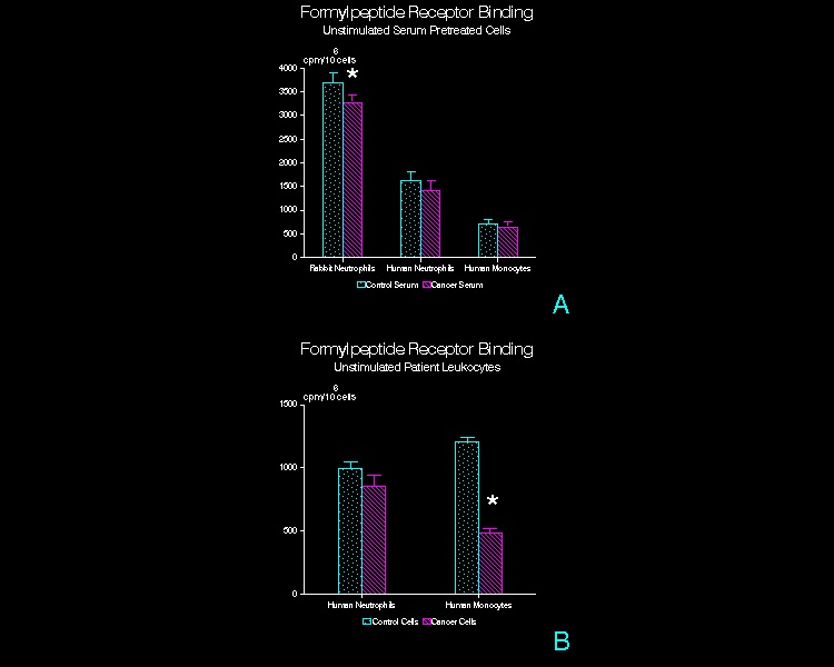
Binding of 3H-FMLP to adherent, unstimulated leukocytes exposed to 3H-FMLP for 60 min at 4°C.
(A)  Serum pretreated normal leukocytes.  Adherent rabbit peritoneal PMN, human peripheral blood PMN, and human peripheral blood mononuclear leukocytes were pre­treated with CT or CA serum prior to exposure to 3H-FMLP.  (n=15, rabbit PMN; n=20, human PMN; n=12, human mononuclear leukocytes; mean + SD)  *  p<0.01  (B)  Patient peripheral blood leukocytes.  PMN and mononuclear leukocytes isolated from venous blood samples obtained from tumor patients or control subjects were exposed to 3H-FMLP at 4°C.  (n=3; mean + SD)  *  p<0.05 


Figure 2
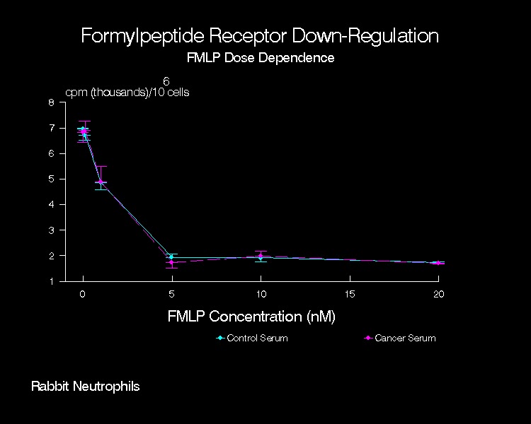
Down-regulation of formylpeptide receptors in the presence of varying concen­trations of unlabeled FMLP by adherent rabbit peritoneal PMN pretreated with CT or CA serum.  Shown is a representative experiment (n=4; mean + SD). 


Figure 3

Time course of formylpeptide receptor down-regulation for adherent rabbit peritoneal PMN pretreated with CT or CA serum and incubated with 10 nM unla­beled FMLP.  Shown is a representative experiment (n=4; mean + SD).


Figure 4
 

Formylpeptide receptor recovery after down-regulation for adherent rabbit peritoneal PMN pretreated with CT or CA serum, exposed to 5 nM unlabeled FMLP, rinsed, and further incubated in buffer at 37°C.  Shown is the percent of cell surface receptors expressed as compared to cell samples not exposed to unla­beled FMLP (i.e., 100% receptor expression)  (n=3; mean + SD).


Figure 5

Uptake of 3H-FMLP (20 nM) at 37°C by adherent rabbit PMN pretreated with CT or CA serum.  Uptake of 3H-FMLP in the CA serum pretreated group is significantly reduced at 10 min (p<0.003), 20 min (p<0.001) and 40 min (p<0.015).  Shown is a representative experiment (n=8).


Figure 6

Uptake of 3H-FMLP at 37°C in the presence of 20 nM 3H-FMLP by adherent normal human leukocytes pretreated with CT or CA serum.  (A)  Uptake in the CA serum pretreated cells is significantly reduced at 40 min (p<0.01) and 60 min (p<0.04).  Shown is a representative experiment (n=6; mean + SD).  (B)  Uptake in CA serum pretreated mononuclear leukocytes is significantly reduced at 40 min (p<0.01) in the CA serum pretreated cells.  Shown is a representative experiment (n=5; mean + SD).  


Figure 7

Uptake of 3H-FMLP at 37°C in the presence of 20 mM 3H-FMLP by adherent PMN isolated from control subjects and tumor patients.  Uptake in the CA patient PMN is significantly reduced at 20 min (p<0.001), 40 min (p<0.001) and 60 min (p<0.002).  Shown is a representative experiment (n=3; mean + SD).

FYI.  The content on this blog is copyright protected by the author.  Feel free to read, copy, and disseminate the studies described here.  When it is appropriate to recognize and attribute the work of others, please indicate the origin of the work by citing this blog URL as an electronic web citation .

Thursday, March 29, 2012

Myosin II and Myosin Light Chain Kinase in Leukocytes from Cancer Patients


March 29, 2012

So, here we go.  The first installment in this series is a manuscript about non-muscle myosins found in human leukocytes.  This paper showed that PMN and monocytes from patients with head and neck cancer contain myosin II and also MLCK.  Myosin levels were decreased in leukocytes from cancer patients compared to those from normal subjects, whereas MLCK levels were similar in PMN and monocytes from both patient groups.  After in vitro exposure to formylpeptide chemoattractant, myosin levels decreased significantly in the PMN and monocytes obtained from normal subjects but were not altered in cancer patient leukocytes.

This suggested that myosin was abnormally regulated in cancer patient leukocytes and that leukocytes from those patients may have been 'pre-activated' in situ.  Such pre-activation might indicate that the host had been actively repulsing an advanced cancer.  However, the long-recognized chemotactic inhibition seen in the leukocytes from such patients suggests just the opposite, that the leukocytes had been activated in situ, had then expended their efforts, had down-regulated their surface receptors, and then became more or less refractile to further stimulation both in situ and in vitro.

Below is the full paper and a link to the PDF:

Myosin II and Myosin Light Chain Kinase in Neutrophils and Monocytes from Cancer Patients


Myosin II and Myosin Light Chain Kinase in

Neutrophils and Monocytes from Cancer Patients
 
by
 
Robert J. Walter, Tzu-Chieh Chao 1, Amelia H. Janeczek 1,

John R. Danielson, and Hernan M. Reyes


Department of Surgery, Cook County Hospital and Hektoen Institute for Medical Research, Chicago, IL

1 Department of Anatomy and Cell Biology, University of Illinois at Chicago, Chicago, IL


Address all correspondence to:
           
Robert J. Walter, Ph.D.           
Department of Surgery
Hektoen Institute for Medical Research
625 South Wood Street
Chicago, IL   60612

Telephone:  (312) 633-7237    
FAX:          (312) 732-3102

Running Title:  Myosin in Tumor Patient Leukocytes

Keywords:   chemotaxis, neutrophils, monocytes, myosin, myosin light chain kinase, cancer,  polarization

Abbreviations

cAMP, 3',5'-cyclic adenosine monophosphate; CT, control; CA, cancer; PMN, polymorphonuclear leukocyte; MLCK, myosin light chain kinase; BSA, bovine serum albumin; FITC, fluorescein isothiocyanate; FMLP, N-formyl-methionyl-leucyl-phenylalanine; HEPES, N-2-hydroxyethylpiperazine-N-2'-ethanesulfonic acid; HBS, Hank's balanced salt solution


ABSTRACT

In vitro chemotaxis of monocytes obtained from tumor patients is often severely depressed but the mechanism of this inhibition is not known.  The actomyosin cytoskeleton is thought to play a pivotal role in generating the forces required for cells to perform activities such as chemotaxis.  This study has employed flow cytometry and fluorescence microscopy to examine two components of the actomyosin machinery, i.e., myosin II and myosin light chain kinase (MLCK) in unstimulated and in formylpeptide-stimulated neutrophils and monocytes isolated from normal (CT) and cancer patient (CA) peripheral blood samples.  Decreased amounts of myosin were observed in unstimulated CA neutrophils and monocytes compared to unstimulated CT leukocytes.  Upon stimulation with formylpeptide chemoattractant, the amount of myosin detected in CT leukocytes decreased markedly but in CA leukocytes was altered very little.  Similar amounts of MLCK were observed in unstimulated CT and CA leukocytes and in formylpeptide-stimulated cells.  In the fluorescence microscope, adherent monocytes and PMN showed diffuse cytoplasmic myosin and MLCK fluorescence throughout the cytoplasm with some increase in intensity at the trailing end (uropod) in formylpeptide-stimulated cells.  Since the actomyosin cytoskeleton is intricately involved in leukocyte chemotaxis, alterations in the cytoskeleton may dramatically affect cell motility.  CA leukocytes exhibited decreased baseline myosin levels and a vastly different response to formylpeptide stimulation as compared to CT leukocytes.  Unstimulated CA monocytes also exhibited increased spontaneous polarization but FMLP-stimulated polarization was inhibited.  These cytoskeletal alterations and changes in the response of CA leukocytes to formylpeptide stimulation may result in decreased chemotaxis by these cells.


INTRODUCTION
           
The actomyosin cytoskeleton is thought to play a prominent role in nonmuscle cell motility.  In leukocytes and single cell eukaryotes such as Dictyostelium discoideum and Acanthamoeba castellanii, cytoplasmic actin and myosin (myosin II or conventional myosin) have been studied extensively and are known to generate the forces that drive chemotaxis (1,2).  Two types of models have been developed to describe the mechanism of force generation in these cells.  The first of these is the actin thin filament-based model which focuses upon the extensive actin remodeling known to occur during chemotactic activation.  Within seconds of exposure to a chemoattractant, extensive actin polymerization occurs followed over the next few minutes by a much slower depolymerization (3,4).  While these events are occurring, actin-rich pseudopods are extended by the cell and, in time, the entire cell becomes polarized structurally with actin concentrated in both the lamellipod and uropod (4-7).  This model proposes that the formation and breakdown of a three dimensional actin lattice is responsible for the cytoplasmic protrusive and contractile activities associated with cell locomotion (2).  The regulation of actin polymerization and depolymerization is thought to involve a range of known actin binding proteins (filamin, acumentin, profilin, gelsolin, etc.) and cytoplasmic calcium (2,8,9). 
           
Myosin II, one of these actin binding proteins, is central in the second model of force generation, the myosin thick filament-based model.  According to this, after chemotactic activation and concurrent with actin remodeling, myosin heavy chain is transiently dephosphorylated and myosin light chain is phosphorylated (10,11) triggering myosin heavy chain assembly into thick filaments (2,12,13).  Subsequently, myosin heavy chain phosphorylation occurs, impeding further assembly of myosin thick filaments (14,15), and leading to depolymerization of existing myosin filaments (16).  During thick filament  assembly in Dictyostelium, myosin shifts in position from the endoplasm into the cortical cytoplasm underlying the plasma membrane and also becomes associated with the posterior uropod (15,17).  In lower eukaryotes and 3T3 fibroblasts, myosin II is largely excluded from the lamellipod during periods of intense membrane ruffling (16,18,19) but in motile leukocytes and HeLa cells, myosin was localized in the lamellipod not the uropod (20).  The reason for this discrepancy is not known.  Nonetheless, when colocalized in the uropod, it is thought that myosin thick filaments may interact with actin by means of myosin ATPase to generate the forces required to drive cell migration and assist in tail retraction (13,21).   These actomyosin interactions are probably regulated by calcium (22) and the phosphorylation of the myosin light chain by myosin light chain kinase (MLCK) (23-25). 

The relative importance of myosins I and II in cell locomotion has been the subject of much recent study.  Genetically altered Dictyostelium deficient in myosin light or heavy chains exhibited markedly decreased rates of locomotion and defective pseudopod formation (26).  In Acanthamoeba, microinjection of monoclonal anti-myosin II slows but does not stop locomotion (19).  Similarly, myosin I deficient Dictyostelium exhibited slowed chemotaxis and decreased phagocytic rates, but no other phenotypic changes (27).  Thus, in lower eukaryotes, it seems likely that both forms of myosin are required for normal cell motility.  However, in vertebrate leukocytes, no role for myosin I can be assigned because it has yet to be demonstrated in these cells (28).  It is presumed that the activation sequence described above is necessary to bring about normal chemotaxis and that alterations in any of the steps of this sequence may result in defective or reduced chemotaxis.  A severe defect in monocyte chemotaxis has been demonstrated in many cancer patients, but the cause of this defect is not known (29,30).  Monocytes, macrophages, and neutrophils are thought to participate directly in tumor destruction (31,32) and studies of experimental animal tumor models have consistently demonstrated that these leukocytes are potent anti-tumor effectors (33).  Monocyte and neutrophil accumulation at sites of tumor growth is accomplished by means of chemotaxis and this response appears to be an essential aspect of the antitumor response (34).  Due to the significance of the leukocyte cytoskeleton in chemotactic activation, we have examined cell polarization as well as the myosin II and MLCK content and distribution in normal leukocytes and in leukocytes from patients with advanced cancer.  As compared to normal leukocytes, unstimulated PMN and monocytes from these patients expressed reduced levels of myosin, monocytes showed reduced cell polarization, and increased spontaneous polarization.  After formylpeptide stimulation, normal PMN and monocytes exhibited reduced myosin levels, but leukocytes from cancer patients exhibited no change in myosin content and reduced cell polarization.  This abnormal modulation of myosin in cancer patient leukocytes may contribute to the chemotactic and polarization defect observed in leukocytes from these patients.


MATERIALS AND METHODS

Selection of Patients

Patients selected for this study were recently diagnosed as having advanced tumors (sarcoma or carcinoma) but were not yet receiving chemotherapy, radiation therapy, or other medications at the time of venipuncture.  Non-cancer patient controls were selected to give age and sex distributions comparable to the cancer patient group. 

Cells and Incubation Conditions

PMN and mononuclear leukocytes were isolated from venous blood collected in EDTA or heparin as described previously (30,35).  Erythrocytes were sedimented at 1xg in 1.25% dextran (pyrogen-free) and the leukocyte-rich plasma drawn off.  Two ml of plasma were diluted 1:1 with a buffer (HBS) containing 140 mM NaCl, 10 mM KCl, 10 mM HEPES, 5mM glucose, 2 mg/ml bovine serum albumin (BSA), pH 7.4 and layered onto one ml of Lymphocyte Separation Medium (Organon Teknika, Durham, NC).  This gradient was then centrifuged at 500xg for 5 min at room temperature, the mononuclear cells at the interface removed, and the PMN pellet resuspended.  PMN were rinsed in HBS containing 2 mM EDTA and contaminating erythrocytes lysed by hypotonic shock.  The mononuclear fraction was diluted with 5 ml of HBS with 2 mM EDTA and 2.5% dextran and centrifuged for 5 min at 500xg.  The platelet-rich supernatant was removed and the dextran centrifugation procedure repeated twice on the resuspended cell pellet.  Cells were resuspended in HBS containing 1 mM MgCl2 and 0.2 mM CaCl2, counted using a hemacytometer, and stored until use at 4ºC.  Differential counts were also performed on these cell preparations. 

Chemotaxis Assays

Cells were tested for their chemotactic capability in multiwell chemotaxis chambers as previously described (30,35).  Briefly, formyl-methionyl-leucyl-phenylalanine (FMLP) over a range of concentrations (10”-11M to 10”-6M) was placed into the lower wells of a 48 well microchemotaxis chamber (Neuroprobe, Cabin John, MD) and purified neutrophils or monocytes were loaded into the upper wells (25 µl; 20,000 cells).  A polyvinylpyrrolidone-free polycarbonate filter (Nucleopore, Pleasanton, CA) with 5 µm pores separated the upper from the lower wells and the chamber was incubated at 37ºC in a humidified incubator for 2 hours.  Samples were run in triplicate and nuclei in 5 (400X) microscope fields from each well were counted.  Monocytes from all tumor patients studied here exhibited a marked chemotaxis defect (65-81% inhibition) compared to non-cancer controls.

Polarization Assay

Patient and control monocytes suspended in HBS/BSA at 37ºC in siliconized tubes were incubated for various times in the presence or absence of FMLP.  At the completion of each incubation, buffered paraformaldehyde was added to a final concentration of 3%, cells were stained with Hoechst 33258 to aid in cell identification, and monocyte polarization determined for 100 cells using light microscopy.  Monocytes were considered to be polarized if they exhibited a change in the normal, rounded morphology such they became elongated or triangular in shape or exhibited asymmetric lamelipodial extension.

Antibodies and Staining Procedures

Cells were exposed to either buffer alone or to FMLP for increasing periods of time (1 to 10 min) at 37ºC.  For PMN, a uniform field of 1 nM FMLP was employed as stimulant whereas 0.1 nM FMLP was used for monocytes.  Antimyosin and anti-MLCK antibodies were the generous gift of Dr. P. deLanerolle (University of Illinois at Chicago) and were used as described previously with some modifications (36).  Unstimulated or FMLP-treated cells were fixed at 4ºC in 2% paraformaldehyde in phosphate buffered saline (PBS), washed, and then lysed in acetone (-20ºC).  After washing in 50% ethanol, autofluorescence and unreacted fixative were quenched using sodium borohydride (1 mg/ml) in 50% ethanol.  Cells were then rinsed in PBS containing 1% normal goat serum and 0.1% BSA and non-specific binding blocked using 5% normal goat serum.  Cells were then reacted with primary antibody for 1 hr at room temperature.  Either rabbit anti-human platelet myosin serum (1:40 dilution) or affinity purified rabbit anti-turkey gizzard MLCK (1:100 dilution) were used as primary antibodies.  Anti-human platelet myosin antibody reacts strongly with mammalian and avian non-muscle myosin and to a lesser degree with mammalian and avian smooth muscle myosin (37).  Polyclonal rabbit anti-turkey gizzard MLCK antibody reacts with MLCK of smooth muscle but not with actin, myosin, tropomyosin, alpha-actinin, or filamin (36).  Subsequent to this incubation, cells were washed and further incubated in FITC-goat anti-rabbit IgG (1:60 dilution; Cappel) for 1 hr.  Cells were then washed using three changes of PBS.  All buffers and antibodies were passed through a 0.22 µm pore size micropore filter before use to remove aggregates.  For flow cytometry, some cell preparations were stained with phycoerythrin-conjugated MY-9 only (Coulter Immunodiagnostics, Hialeah, FL).   MY-9 is a monoclonal antibody that binds to normal peripheral blood monocytes but not to granulocytes, erythrocytes, platelets, or lymphocytes (38). 


Flow Cytometry

Stained cells were filtered through nylon mesh (25 µm pore size; TETKO Inc., Elmsford, NY) and analyzed using an EPICS C flow cytometer (Coulter Electronics, Hialeah, FL) equipped with a 500 mW argon ion laser emitting at 488 nm.  Forward-angle and right-angle light scatter, fluorescein (535 nm), and phycoerythrin (580 nm) fluorescence were acquired for at least 5000 cells in each experimental group.  Fluorescence was collected using a three-decade logarithmic amplifier.

Some cells were fixed in 2% buffered paraformaldehyde and stained with either phycoerythrin-conjugated MY-9 (Coulter Immunology, Hialeah, FL; diluted 1:30) or with phycoerythrin-conjugated nonspecific mouse immunoglobulin (diluted 1:30).   MY-9 was used to aid in determining the exact monocyte and neutrophil gate settings and to ascertain the proportion of monocytes counted in the monocyte gate. 


Polarization Assay

The time course and concentration dependence of the monocyte polarization response was assessed for monocytes suspended in HBS in siliconized tubes.  Cells were either exposed to a range of concentrations of FMLP for 20 min at 37ºC, to 1 nM FMLP for 0 to 60 min, or to buffer alone for 0 to 60 min.  Incubations were terminated by the addition of paraformaldehyde to a final concentration of 4%, and cells were stained with Hoechst 33258.  Monocytes were identified by nuclear morphology and size using fluorescence microscopy and cell polarization evaluated for 100 cells at each data point.  Cells were considered to be polarized if they exhibited a distinct shape change including triangular shape, asymmetric protuberance formation, or lamellipodial extension.


Fluorescence Microscopy and Photography

For fluorescence microscopy, purified PMN and monocytes were remixed and allowed to adhere to clean glass coverslips at 37ºC for 5 min.  After fixation, extraction, and staining as described above, labeled leukocytes were further stained with Hoechst 33258 (10 µg/ml) in PBS for 10 min at room temperature.  After washing, cover slips were mounted on slides in PBS with 90% glycerol containing paraphenylenediamine (1 mg/ml) (39).  These preparations were examined using a Nikon Optiphot microscope with a Leitz 100 W mercury epiiluminator and photographed using Kodak Tri-X Pan (ASA 400) or Ektachrome (ASA 1600).


Statistical Evaluation of Data

Mean channel numbers for each time or treatment were treated as individual parametric values and groups of these values were compared using unpaired t-tests.  Chi-square was employed to evaluate non-parametric cell polarization data.  Mean channel numbers are the average of three separate experiments (mean ± SD) performed on blood samples from different CT and CA subjects as starting material.  A probability value < 0.05 was considered significant.


RESULTS

Myosin Redistributes into the Uropod upon FMLP Stimulation

Unstimulated, adherent CT leukocytes were generally uniformly spread except for a few cells (< 5%) that were spontaneously polarized.  Antimyosin staining disclosed diffuse cytoplasmic fluorescence in both rounded and spontaneously polarized cells (Figure 1a).  Increased amounts of fluorescence were seen in the central regions of the cells but very little fluorescence was associated with the cortical regions of the cell.  Unstimulated PMN and monocytes from CA patients were similar in overall appearance to CT leukocytes, but exhibited somewhat decreased levels of fluorescence (Figures 1b, c).  In all groups, monocytes displayed less intense fluorescence than did PMN.

Formylpeptide-stimulated CT leukocytes were structurally polarized exhibiting a distinct leading lamellipod and trailing uropod (Figures 1d-g).  Antimyosin staining was more concentrated beneath the plasma membrane than that seen in unstimulated cells.  PMN showed distinct concentrations of fluorescence in the trailing uropod region of polarized cells and monocytes showed slight increases in the the amount of fluorescence in the uropod.  Formylpeptide-stimulated PMN and monocytes from CA patients (Figure 1f, g) were similar in overall appearance to stimulated CT leukocytes (Figure 1d, e), but in general exhibited decreased levels of fluorescence.  Differences in fluorescence intensity were clearly evident in specimens examined in the flow cytometer.


MLCK is Diffusely Distributed

Unstimulated, adherent CT and CA PMN and monocytes stained for MLCK exhibited faint diffuse cytoplasmic fluorescence (Figures 2a-d).  Formylpeptide-stimulated CT and CA leukocytes displayed MLCK staining that was in most cases diffuse throughout the cytoplasm, but in some cells was unmistakably concentrated in the uropod region (Figures 2e, f). 

Myosin Staining Differs in CT and CA Leukocytes

Phycoerythrin-conjugated MY-9 staining was used to confirm the location and cell type found within the monocyte gate (Figure 3).  This was necessary because the extraction protocol used prior to staining for myosin or MLCK resulted in less discrete monocyte distributions as seen in scatterplots (right angle versus forward light scatter).  MY-9 staining of the gated monocyte population revealed that 85% of the cells were monocytes.  Due to their low cytoplasmic granularity, the remainder were presumed to be large lymphocytes.  This same proportion of monocytes was seen in both CA and CT samples. 

Unstimulated CT PMN stained for myosin were seen as discreet unimodal distributions.  As a group (Table 1), unstimulated CT PMN had an average mean channel number of 80 ± 4 (Figure 4) whereas unstimulated PMN from CA patients had a mean channel number of 48 ± 9 (p < 0.005 compared to unstimulated CT).  Upon exposure to 1 nM FMLP, the mean channel number observed for CT PMN declined to 63 ± 5 after 1 minute of exposure and to 61 ± 5 after 4 minutes of exposure to FMLP.  In contrast, under these same conditions the mean channel number for CA PMN increased to 49 ± 10 and 53 ± 11, respectively.

Unstimulated CT monocytes stained for myosin were seen as broad unimodal distributions having a mean channel number of 77 ± 4 whereas unstimulated monocytes from CA patients were seen as broad unimodal distributions having an average mean channel number of 22 ± 6.  Upon exposure to 0.1 nM FMLP, the mean channel number observed for CT monocytes declined to 47 ± 3 after 1 minute of exposure and to 30 ± 5 after 4 minutes of exposure to FMLP.  Under these same conditions the mean channel number for CA monocytes remained virtually unchanged at 21 ± 5 and 20 ± 3, respectively.


MLCK Staining is Similar in CT and CA Leukocytes

Unstimulated CT PMN stained for MLCK (Table 2) were seen as a unimodal distributions having a mean channel number of 63 ± 10, whereas unstimulated PMN from CA patients displayed mean channel numbers of 62 ±10.  Upon exposure to 1 nM FMLP, the mean channel number observed for CT PMN was 67 ± 14 after 1 minute of exposure to FMLP and 64 ± 17 after 4 minutes.  Under these same conditions, the mean channel number for CA PMN increased slightly to 63 ±10 and 65 ±10, respectively. 

Unstimulated CT monocytes stained for MLCK were seen as a broad unimodal peak having a mean channel number of 61 ± 3 whereas unstimulated monocytes from CA patients exhibited unimodal peaks with an average mean channel number of 55 ± 3.  Upon exposure to 0.1 nM FMLP, the mean channel number observed for CT monocytes increased to 70 ± 3 and 72 ± 2 after 1 and 4 minutes of exposure to FMLP, respectively.  Under these same conditions, the mean channel number for CA monocytes increased to 62 ± 5 and 65 ± 3, respectively.


Spontaneous and FMLP-Stimulated Cell Polarization is Altered in CA Monocytes 

CT and CA patient monocytes were exposed to buffer alone or to different concentrations of FMLP for 20 min at 37ºC (Figure 5).  In the presence of FMLP, polarization was significantly reduced in CA as compared to CT monocytes with maximal polarization evident at 10-9 M FMLP.  However, in buffer alone, polarization was significantly increased in CA as compared to CT monocytes.
           
As seen in Figure 6, CT and CA patient monocytes were incubated with 10-9 M FMLP or in buffer alone for times ranging from 0 to 60 min.  In buffer alone, the fraction of polarized CA monocytes was significantly greater at all times (except 0 time) than that seen for CT monocytes.  FMLP-stimulated CA monocytes exhibited significantly decreased polarization for incubation times between 5 and 30 min but significantly increased polarization after 60 min of incubation as compared to that seen for CT monocytes.


DISCUSSION    

We have examined myosin staining, MLCK staining, and cell polarization in unstimulated and formylpeptide-stimulated PMN and monocytes isolated from CT and CA patients.  We have observed:  1)  decreased myosin staining in PMN and monocytes from CA patients as compared to leukocytes taken from CT patients, 2)  that stimulation with formylpeptide decreases myosin staining in CT leukocytes but not in leukocytes from CA patients, 3)  comparable levels of MLCK in CT and CA leukocytes, and similar levels of MLCK staining in both cell groups when stimulated with formylpeptide, 4)  that unstimulated CA monocytes exhibited increased polarization but that formylpeptide-stimulated polarization was inhibited as compared to CT monocytes.

Myosin staining of monocytes (CT and CA) was less intense than that of PMN (CT and CA) as seen in the fluorescence microscope and in the flow cytometer.  After formylpeptide stimulation of CT patient leukocytes, distinct morphological alterations became evident.  Myosin staining became more concentrated beneath the plasma membrane especially in the uropod region and markedly reduced amounts of myosin staining were seen.  Amounts of MLCK staining remained unchanged but staining was sometimes concentrated in the uropod.  Previous reports on the distribution of myosin in chemotactically-activated cells are somewhat contradictory and there have been few reports on myosin distributions in higher eukaryotic cells such as leukocytes.  Most of the recent studies in lower eukaryotes have shown myosin II within cellular protrusions or in the endoplasm but have not described its transcellular location (anterior, middle, or posterior) in the motile cell. 

In unstimulated cells (see Table 3), myosin II is found diffusely distributed in the endoplasm and often absent from the cell cortex.  In chemotactically activated amoebae, myosin II is consistently found in the uropod and the cortical cytoplasm.  Similarly, myosin II co-caps with surface Ig on human lymphocytes to a region analogous to the uropod of a motile cell and is absent or depleted from the leading edge of carcinoma cells.  Using fluorescence photobleaching recovery, DeBiasio et al. (40) have also found that myosin is present but immobile in the leading edge of motile 3T3 cells.  However, in rabbit PMN responding to a gradient of complement fragments, myosin II was localized to the lamellipod (20).  Our findings differ from those of Valerius et al. in that we find myosin most often in the uropod of human PMN exposed to FMLP.  The contradiction may be due to differences in cell types, species, chemoattractants, or methods of applying the attractant (yeast-generated gradient versus uniform field).  However, our findings concur closely with those obtained with lower eukaryotes, lymphocytes, and cultured fibroblasts and therefore serve to clarify our understanding of myosin distribution in a range of motile cell types.

The quantitative changes in myosin levels observed here during leukocyte activation, i.e. decreases in myosin staining, were somewhat unexpected in view of some previous reports.  White et al. (41) have shown for rabbit PMN that the levels of myosin associated with the Triton-insoluble cytoskeleton remained unchanged when they were assessed 15 seconds after FMLP stimulation and Feinstein et al. (42) have shown a substantial thrombin-induced increase in the myosin content of the Triton-insoluble platelet cytoskeleton.  However, the present study has utilized preparative and analytic procedures that differ considerably from those employed in the aforementioned studies.  Most notably the time of exposure to FMLP was greater here (1 and 4 min) as compared to the study of White et al. (15 seconds) and the fixation/extraction procedures employed here involved chemical fixatives and organic solvents (ethanol, acetone) rather than detergents as were used by others.  On the other hand, Dharmawardhare et al. (16) have shown that the myosin II content of the Dictyostelium cytoskeleton peaks at 25-30 seconds after cAMP stimulation and reaches a minimum, about 50% below baseline levels, 40 seconds after cAMP stimulation.  Levels of cytoskeleton-associated myosin remained significantly below baseline throughout the remainder of the 70 second time course studied there.  The myosin content of human leukocytes seen in the present study at both 1 and 4 minutes after FMLP stimulation was correspondingly low.

In Acanthamoeba, Baines and Korn (43) have shown that plasma membrane associated myosin IC is labile to saponin treatment, whereas contractile vacuole associated myosin IC is preserved.  Thus, it seems that myosin I may be more or less extractable depending on its subcellular location.  Baines and Korn have suggested that this differential vulnerability to extraction may be related to the extent to which myosin interacts with membrane lipids.  In this study, we are localizing and quantitating myosin II, not myosin IC, but the principle of differential extractability or solubility may also apply here.  Such differential extractability may be related to putative interactions between myosin II and membrane lipids or to the state of myosin phosphorylation.  When myosin heavy chain is phosphorylated, myosin thick filaments depolymerize, and myosin may be lost from the cytoskeleton (16,44).  Chemoattractant stimulation of leukocytes from CT patients may cause myosin heavy chain to be phosphorylated or otherwise redistributed and less tightly associated with the cell constituents that are preserved under the fixation/extraction conditions used here.
We have also observed that MLCK expression remained relatively unchanged after FMLP stimulation.  This suggests that the markedly decreased myosin staining observed after FMLP stimulation was not simply the result of increased nonspecific protein extraction during specimen preparation.  The decrease in myosin staining seen in CT PMN and monocytes upon FMLP exposure appears to be selective for myosin. 

In unstimulated leukocytes from CA patients, we have observed dramatically reduced myosin staining compared to that seen in unstimulated leukocytes from CT patients.  On the other hand, myosin in CA leukocytes was virtually unaffected by exposure to FMLP such that these cells did not exhibit the expected decrease in myosin staining upon FMLP stimulation.  Since myosin phosphorylation apparently determines the extent of the association between myosin and the cytoskeleton, these findings suggest that myosin phosphorylation/ dephosphorylation may be abnormal in CA leukocytes.  We hypothesize that leukocytes from CA patients may be pre-activated by exposure to circulating factors present in the blood and this may reduce cytoskeleton-associated myosin even before in vitro stimulation with FMLP.  Upon addition of FMLP, myosin levels remain unchanged because cells had become refractory to further stimulation. 

As further evidence of this, CA monocyte samples not pretreated with FMLP (i.e., unstimulated) exhibit increased numbers of polarized cells as compared to unstimulated CT monocytes.  Furthermore, CA monocyte samples stimulated by FMLP exhibit decreased numbers of polarized cells at all times less than 40 min and at all FMLP concentrations used here.  Others have studied cell polarization in normal monocytes treated with CA patient effusions (45), tumor-derived low molecular weight factors (46), and fragments of retroviral p15E peptide analogues such as the peptide LDLLFL (47), but little data on polarization in CA patient leukocytes has been reported.  Results from previous studies are generally in agreement with those described here.  Interestingly, Cianciolo et al. (45) showed that cancer patient pleural or peritoneal effusions contained high molecular weight factors that were stimulatory and low molecular weight factors that were inhibitory to normal monocyte polarization.  The data presented here suggest that similar effects may be evident in leukocytes isolated from CA patients.  The high levels of polarization evident in unstimulated CA monocytes indicate that these cells are pre-activated as they are obtained from the patient.  This further suggests that the reductions in cell polarization seen in FMLP-stimulated CA monocytes may result from desensitization of these cells toward FMLP.

The cellular mechanism of the chemotactic defect in CA patient monocytes is unknown.  However, it is thought that a serum-borne cell-directed inhibitor may be responsible for the defect (48,49).  This inhibitor causes decreased monocyte polarization in response to chemoattractant (50,51) [unpublished data], alterations in formylpeptide receptor expression (35), suppression of the respiratory burst (52), and inhibition of protein kinase C-related cell functions (53).  Myosin heavy and light chains are known substrates for protein kinase C (44), but the role of protein kinase C in regulating myosin phosphorylation in non-muscle cells is not known (Wilson and DeLan, 1992).  The alterations in myosin content of leukocytes in CA patients described here may be directly involved in the inhibition of cell polarization and the chemotactic deficiency seen in cells from these patients.  This may contribute to the inability of these immune effectors to deter the growth and spread of neoplasia and to the susceptibility of these patients to life-threatening bacterial infections (54).

ACKNOWLEDGMENTS

The authors would like to thank Dr. Abraham Mark and Robert Novak for their assistance with flow cytometry and John Krewer for technical assistance.  This work was supported in part by the American Cancer Society, Illinois Division. 


REFERENCES

 1. DEVREOTES PN, ZIGMOND SH. Chemotaxis in eukaryotic cells:  A focus on leukocytes and Dictyostelium. Ann Rev Cell Biol 1988;4:1054-1058.

 2. SOUTHWICK FS, STOSSEL TP. Contractile proteins in leukocyte function. Sem Hematol 1983;20:305-321.

 3. WALLACE PJ, WERSTO RP, PACKMAN CH, et al. Chemotactic peptide-induced changes in neutrophil actin conformation. J Cell Biol 1984;99:1060-1065.

 4. HOWARD TH, ORESAJO CO. The kinetics of chemotactic peptide-induced changes in F-actin content, F-actin distribution, and the shape of neutrophils. J Cell Biol 1985;101:1078-1085.

 5. YUMURA S, MORI H, FUKUI Y. Localization of actin and myosin for the study of ameboid movement in Dictyostelium using improved immunofluorescence. J Cell Biol 1984;99:894-899.

 6. OLIVER JM, KRAWIEC JA, BECKER EL. The distribution of actin during chemotaxis in rabbit neutrophils. J Reticuloend Soc 1978;24:697-704.

 7. SULLIVAN JA, MANDELL GL. Motility of human polymorphonuclear neutrophils:  Microscopic analysis of substrate adhesion an distribution of F-actin. Cell Motil 1983;3:31-46.

 8. STOSSEL TP, HARTWIG JH, YIN HL, et al. The motor of leukocytes. Fed Proc 1984;43:2760-2763.

 9. WEEDS A. Actin binding proteins:  Regulators of cell architecture and motility. Nature 1982;296:811-816.

10. BERLOT CH, SPUDICH JA, DEVREOTES PN. Chemoattractant-elicited increases in myosin phosphorylation in Dictyostelium. Cell 1985;43:307-314.

11. FECHHEIMER M, ZIGMOND SH. Changes in cytoskeletal proteins of polymorphonuclear leukocytes induced by chemotactic peptides. Cell Motil 1983;3:349-361.

12. IKEBE M, INAGACKI M, NAKA M, et al. Correlation of conformation and phosphorylation and dephosphorylation of smooth muscle myosin. J Biol Chem 1988;263:10698-10704.

13. CRAIG R, SMITH R, KENDRICK-JONES J. Light chain phosphorylation controls the conformation of vertebrate non-muscle and smooth muscle myosin molecules. Nature 1983;302:436-439.

14. SPUDICH JA. In pursuit of myosin function. Cell Regul 1989;1:1-11.

15. BERLOT CH, DEVREOTES PN, SPUDICH JA. Chemoattractant-elicited increases in Dictyostelium myosin phosphorylation are due to changes in myosin localization and increases in kinase activity. J Biol Chem 1987;262:3918-3926.

16. DHARMAWARDHANE S, WARREN V, HALL AL, et al. Changes in the association of actin-binding proteins with the actin cytoskeleton during chemotactic stimulation of Dictyostelium discoideum. Cell Motil Cytoskel 1989;13:57-63.

17. YUMURA S, FUKUI Y. Reversible cyclic AMP-dependent change in distribution of myosin thick filaments in Dictyostelium. Nature 1985;314:194-196.

18. HONER B, CITI S, KENDRICK-JONES J, et al. Modulation of cellular morphology and locomotory activity by antibodies against myosin. J Cell Biol 1988;107:2181-2189.

19. SINARD JH, POLLARD TD. Microinjection into Acanthamoeba castellanii of monoclonal antibodies to myosin-II slows but does not stop cell locomotion. Cell Motil Cytoskel 1989;12:42-52.

20. VALERIUS NH, STENDAHL O, HARTWIG JH, et al. Distribution of actin-binding protein and myosin in polymorphonuclear leukocytes during locomotion and phagocytosis. Cell 1981;24:195-202.

21. CHEN WT. Mechanism of retraction of the trailing edge during fibroblast movement. J Cell Biol 1981;90:187-200.

22. HOLZPAFEL G, WEHLAND J, WEBER K. Calcium control of actin-myosin based contraction in Triton models of mouse 3T3 fibroblasts is mediated by the myosin light chain kinase (MLCK)-calmodulin complex. Exptl Cell Res 1983;148:117-126.

23. WILSON AK, GORGAS G, CLAYPOOL WD, et al. An increase or a decrease in myosin II phosphorylation inhibits macrophage motility. J Cell Biol 1991;114:277-283.

24. ADELSTEIN RS. Regulation of contractile proteins by phosphorylation. J Clin Inv 1983;72:1863-1866.

25. CANDE WZ, EZZELL RM. Evidence for regulation of lamellipodia and tail contraction in glycerinated chicken embryonic fibroblasts by myosin light chain kinase. Cell Motil 1986;6:640-648.

26. WESSELS D, SOLL ,D.R., KNECHT D, et al. Cell motility and chemotaxis in Dictyostelium amebae lacking myosin heavy chain. Devel Biol 1988;128:164-177.

27. JUNG G, HAMMER JA. Generation and characterization of Dictyostelium cells deficient in a myosin I heavy chain isoform. J Cell Biol 1990;110:1955-1964.

28. WILSON AK, POLLENZ RS, CHISHOLM RL, et al. The role of myosin I and II in cell motility. Canc Metas Revs 1992;11:79-91.

29. WALTER RJ, DANIELSON JR, VAN ALTEN PJ, et al. Defects in monocyte chemotaxis in patients with neoplastic disease. J Surg Res 1986;41:215-224.

30. WALTER RJ, DANIELSON JR. Defective monocyte chemotaxis in patients with epidermoid tumors of the head and neck. Arch Otolar 1985;111:538-540.

31. HIBBS JB, LAMBERT LH, REMINGTON JS. Possible role of macrophage mediated nonspecific cytotoxicity in tumour resistance. Nature N B 1972;235:48-50.

32. MORTON DL. Horizons in tumor immunology. Surgery 1973;74:69-79.

33. NORTH RJ, KIRSTEN DP, TUTTLE RL. Subversion of host defense mechanisms by murine tumors.  I.  A circulating factor that suppresses macrophage-mediated resistance to infection. J Exptl Med 1976;143:559-573.

34. EVANS R. Macrophages in syngeneic animal tumors. Transpl 1972;14:468-473.

35. WALTER RJ, DANIELSON JR. Characterization of formylpeptide chemoattractant binding on neutrophils and monocytes from patients with head and neck cancer. J Natl Canc Inst 1987;78:61-69.

36. DELANEROLLE P, ADELSTEIN RS, FERAMISCO JR, et al. Characterization of antibodies to smooth muscle myosin kinase and their use in localizing myosin kinase in nonmuscle cells. Proc Natl Acad Sci 1981;78:4738-4742.

37. KAWAMOTO S, ADELSTEIN RS. Characterization of myosin heavy chains in cultured aorta smooth muscle cells:  A comparative study. J Biol Chem 1987;262:7282-7288.

38. GRIFFIN JD, LYNCH D, SABBATH K, et al. A monoclonal antibody reactive with normal and leukemic human myeloid progenitor cells. Leuk Res 1984;8:521-529.

39. JOHNSON GD, NOGUEIRA ARAUJO GM. A simple method of reducing the fading of immunofluorescence during microscopy. J Immunol Meth 1981;43:349-350.

40. DEBIASIO RL, WANG L-L, FISHER GW, et al. The dynamic distribution of fluorescent analogues of actin and myosin in protrusions at the leading edge of migrating Swiss 3T3 fibroblasts. J Cell Biol 1988;107:2631-2645.

41. WHITE JR, NACCACHE PH, SHA'AFI RI. Stimulation by chemotactic factor of actin association with the cytoskeleton in rabbit neutrophils. J Biol Chem 1983;258:14041-14047.

42. FEINSTEIN MB, EGAN JJ, OPAS EE. Reversal of thrombin-induced myosin phosphorylation and the assembly of cytoskeletal structures in platelets by the adenylate cyclase stimulants prostaglandin D2 and forskolin. J Biol Chem 1983;258:1260-1267.

43. BAINES IC, KORN ED. Localization of myosin IC and myosin II in Acanthamoeba castellanii by indirect immunfluorescence and immunogold electron microscopy. J Cell Biol 1990;111:1895-1904.

44. KELLEY CA, KAWAMOTO ,S., CONTI MA, et al. Phosphorylation of vertebrate smooth muscle and nonmuscle myosin heavy chains in vitro and in intact cells. J Cell Sci 1991;Suppl. 14:49-54.

45. CIANCIOLO GJ, HUNTER J, SILVA J, et al. Inhibitors of monocyte responses to chemotaxins are present in human cancerous effusions and react with monoclonal antibodies to the p15E structural protein of retroviruses. J Clin Inv 1981;68:831-844.

46. TAN IB, BALM FJM, SNOW GB, et al. Immunosuppressive retrovirus-related factors in sera of patients with head and neck cancer. Eur Arch Otolaryn 1990;247:387-390.

47. OOSTENDORP RAJ, SCHAAPER WMM, POST J, et al. Synthetic hexapeptides derived from the transmembrane envelope proteins of retroviruses suppress N-formylpeptide-induced monocyte polarization. J Leuk Biol 1992;51:282-288.

48. CIANCIOLO GJ, MATTHEWS TJ, BOLOGNESI DP, et al. Macrophage accumulation in mice is inhibited by low molecular weight products from murine leukemia viruses. J Immunol 1980;124:2900-2905.

49. MADERAZO EG, ANTON TF, WARD PA. Serum-associated inhibitor of leukocytosis in humans with cancer. Clin Immunol Immunopath 1978;9:166-176.

50. TAN IB, DREXHAGE HA, SCHEPER RJ, et al. Immunosuppressive retroviral p15E-related factors in head and neck carcinomas. Arch Otolaryngol HN Surg 1986;112:942-945.

51. Cianciolo, G.J. and Snyderman, R. Characterisation of an inhibitor of monocyte function in effusions of cancer patients. In: Lymphokines and thymic hormones:  their potential utilization in cancer therapeutics, edited by Goldstein, A.L. and Chirigos, M.A. New York: Raven Press, 1981, p. 205-213.

52. HARRELL RA, CIANCIOLO GJ, COPELAND TD, et al. Suppression of the respiratory burst of human monocytes by a synthetic peptide homologous to envelope proteins of human and animal retroviruses. J Immunol 1986;136:3517-3520.

53. GOTTLIEB RA, KLEINERMAN ES, O'BRIAN CA, et al. Inhibition of protein kinase C by a peptide conjugate bound to a domain of  the retroviral protein p15E. J Immunol 1990;145:2566-2570.

54. INAGACKI J, RODRIGUEZ V, BODEY GP. Causes of death in cancer patients. Cancer 1974;33:568-578.

55. FUKUI Y, MURRAY J, RIDDELLE KS, et al. Cell behavior and actomyosin organization in Dictyostelium during substrate exploration. Cell Struc Func 1991;16:289-301.

56. HAGEN SJ, KIEHART DP, KAISER DA, et al. Characterization of monoclonal antibodies to Acanthamoeba myosin-I that cross-react with myosin-II and low molecular mass nuclear proteins. J Cell Biol 1986;103:2121-2128.

57. JOCKUSCH RM, HAEMMERLI G, IN ALBON A. Cytoskeletal Organization in locomoting cells of the V2 rabbit carcinoma. Exptl Cell Res 1983;144:251-263.

58. UEDA T, SHIBATA N. Distribution patterns of actin and myosin during leucocyte movement and phagocytosis. Acta Haematol 1983;46:1451-1461.

59. RUBINO S, FIGHETTI M, UNGER E, et al. Location of actin, myosin, and microtubular structures during directed locomotion of Dictyostelium amebae. J Cell Biol 1984;98:382-390.

60. FUKUI Y, LYNCH TJ, BRZESKA H, et al. Myosin I is located at the leading edges of locomoting Dictyostelium amoebae. Nature 1989;341:328-331.

61. SCHREINER GF, FUJIWARA K, POLLARD TD, et al. Redistribution of myosin accompanying capping of surface Ig. J Exptl Med 1977;145:1393-1398.

62. BRAUN J, FUJIWARA K, POLLARD TD, et al. Two distinct mechanisms for redistribution of lymphocyte surface macromolecules.  I.  Relationship to cytoplasmic myosin. J Cell Biol 1978;79:409-418.

63. BOURGUIGNON LYW, SINGER SJ. Transmembrane interactions and the mechanism of capping of surface receptors by their specific ligands. Proc Natl Acad Sci 1977;74:5031-5035.


FIGURES


  Figure 1

Leukocytes were fixed, lysed in acetone, quenched in sodium borohydride, and nonspecific binding blocked with BSA and NGS.  Cells were exposed to rabbit anti-chicken gizzard myosin serum, FITC-labeled goat anti-rabbit IgG, and stained with Hoechst 33258 to aid in the identification of monocytes.  (a)  unstimulated, adherent CT leukocytes showed diffuse cytoplasmic fluorescence in both rounded and spontaneously polarized (arrow) cells.  Very little fluorescence was associated with the plasma membrane.  (b, c)  Unstimulated PMN and monocytes from CA patient.  Nuclear fluorescence (c) clearly distinguishes monocytes from PMN (arrowhead).  Monocytes usually displayed less intense, more diffuse fluorescence than did PMN (d-g).  Formylpeptide-stimulated CT PMN and monocytes exhibited a distinct leading lamellipod and trailing uropod (d,e; arrowheads).  Formylpeptide-stimulated PMN and monocytes from CA patients (f,g).   Antimyosin staining was concentrated beneath the plasma membrane for both PMN and monocytes and prominently in the trailing uropod region of polarized PMN. (a) Magnification bar = 20 µm; (b-g) Magnification bar = 10 µm.


Figure 2

Leukocytes were fixed, lysed in acetone, quenched in sodium borohydride, and nonspecific binding blocked with BSA and NGS.  Cells were exposed to affinity-purified rabbit anti-turkey gizzard MLCK, FITC-labeled goat anti-rabbit IgG, and stained with Hoechst 33258 to aid in the identification of monocytes.  Unstimulated, adherent CT (a, b) and CA (c, d) PMN and monocytes exhibited faint diffuse cytoplasmic fluorescence.  Formylpeptide-stimulated CT (e) and CA (f) leukocytes displayed MLCK staining that was in most cases diffuse throughout the cytoplasm, but in some cells (arrows) was concentrated in the uropod region.  (a-d) Magnification bar = 10 µm; (e, f) magnification bar = 20 µm.


Figure 3

Right angle versus forward light scatter (a) of CT PMN, monocytes, and lymphocytes.  Monocytes were enriched, fixed, extracted with acetone, treated with antimyosin followed by FITC-conjugated goat antirabbit IgG and finally with phycoerythrin-conjugated MY-9.  The upper right gate surrounds the region containing residual PMN contaminating the monocyte suspension.  Due to their distinct granularity and size this region is easily distinguished in most preparations.  The lower left gate surrounds the region typically occupied by monocytes.  Lymphocytes, red blood cells, and platelets are seen to the far left.  MY-9 staining (b) of the myosin-positive cells found in this gate indicated that 85% of the cells observed were monocytes.


Figure 4

Right angle versus forward light scatter (A), a representative plot showing purified CT PMN gate and cells.  Cells were fixed, extracted, and stained for myosin.  Log green fluorescence versus cell count histograms (B) showed a sharp, unimodal distribution having a mean channel number (for this sample) of 78 ± 9 (SD).


Figure 5

CT (filled circles) and CA (open circles) patient monocytes were exposed to buffer alone or to different concentrations of FMLP for 20 min at 37ºC.  At each concentration, monocyte polarization in CA samples was significantly different (p<0.01) than that seen in CT samples.  Data are expressed as mean ± SD (n = 6).

 Figure 6

CT (filled circles) and CA (open circles) patient monocytes were incubated with 10-9 M FMLP (solid lines) or in buffer alone (dotted lines) for times varying from 0 to 60 min.  In buffer alone, the fraction of polarized CA monocytes was significantly greater (p<0.05) at all times (except 0 time) than that seen for CT monocytes.  FMLP-stimulated CA monocytes exhibited significantly decreased (p<0.05) polarization for incubation times between 5 and 30 min but significantly increased (p<0.05) polarization after 60 min of incubation as compared to that seen for CT monocytes.  Data are expressed as mean ± SD (n = 6).


TABLE 1


Averaged Mean Channel Numbers for PMN and Monocytes
from CT and CA Subjects Anti-myosin Staining


PMN                           MONOCYTES

CT           CA               CT           CA
_______________________________________________________________________

      Unstimulated                       80 ± 4       a 48 ± 9        77 ± 16      a 22 ± 6

      FMLP-treated

                  1 min                      63 ± 5       b 49 ± 10       47 ± 3       b 21 ± 5

                  4 min                      61 ± 5         53 ± 11       30 ± 5       b 20 ± 3

_______________________________________________________________________

Leukocytes from 3 CT and 3 CA subjects were studied.  The mean channel numbers for each group were averaged (mean ± SD).

a p<0.005  when compared to unstimulated CT group.

b p<0.05  when compared to FMLP stimulated CT group.


TABLE 2


Averaged Mean Channel Numbers for PMN and Monocytes
from CT and CA Subjects Anti-MLCK Staining


PMN                           MONOCYTES

CT           CA             CT           CA

_______________________________________________________________________


      Unstimulated                       63 ± 10      a 62 ± 10       61 ± 3       a 55 ± 3

      FMLP-treated

                        1 min                67 ± 14      a 63 ± 13       70 ± 3       a  62 ± 5

                        4 min                64 ± 17      a 65 ± 10      b 72 ± 2      ab 65 ± 3


_______________________________________________________________________


Leukocytes from 3 CT and 3 CA subjects were studied.  The mean channel numbers for each group were averaged (mean ± SD).

a Not significantly different from paired CT group.

b p<0.05 when compared to unstimulated group.


TABLE 3

Myosin II Localization in Unstimulated and Chemotactically Activated Cells


Cell type                                             Myosin II distribution            Stimulant        Reference

Dictyostelium discoideum                   excluded from cortex                None                (55)

Dictyostelium discoideum                   rods in endoplasm                     None                (17)

Acanthamoeba castellanii                   cell cortex                                 None                (43)

Acanthamoeba castellanii                   diffuse in cytoplasm                   None                (56)
   
V2 rabbit carcinoma                             absent from lamellipod               None                (57)

Rabbit PMN                                        diffuse                                       None                (20)

Human PMN                                        diffuse                                       None                (58)

Dictyostelium discoideum                   rods in ectoplasm                       cAMP              (17)

Dictyostelium discoideum                   posterior cortex                         cAMP                (5)

Dictyostelium discoideum                   uropod only                               cAMP              (59)

Dictyostelium discoideum                   uropod only                               cAMP              (60)

3T3 fibroblasts                                     absent from protrusions              Wound             (40)
                                                            immobile in lamellipod     

Human lymphocytes                              uropod                                      Ig capping        (61)

Human lymphocytes                              uropod                                      capping            (62)

Mouse T lymphocyte                            uropod                                      capping            (63)

Rabbit PMN, HeLa cells                      lamellipod                                  complement      (20)


FYI.  The content on this blog is copyright protected © by the author.  Feel free to read, copy, and disseminate the studies described here.  When it is appropriate to recognize and attribute the work of others, please indicate the origin of the work by citing this blog URL as an electronic web citation .